Dental Case: Problems

Multiple Decayed Lower Molars With Abscess Risk

Severity: 

Multiple Decayed Lower Molars With High Risk of Abscess Case Analysis

What Is Seen in This Case

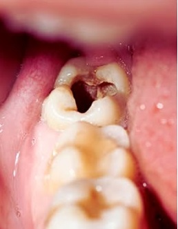
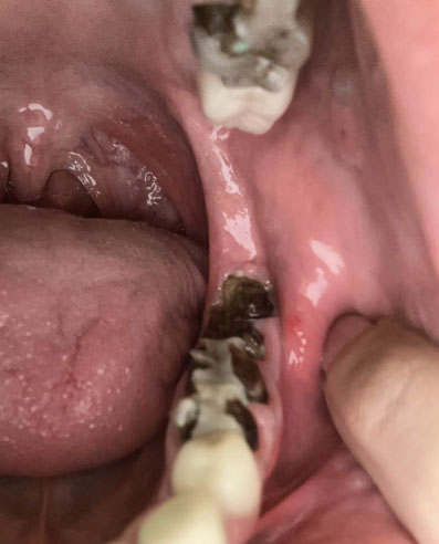
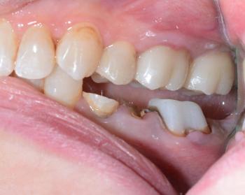
The image shows the lower jaw (mandible) with multiple back molars affected by visible decay. On both the left and right sides, at least one molar shows brown cavitated lesions, indicating advanced tooth decay. The surrounding gum tissue appears slightly irritated, and the floor of the mouth is clearly visible.

This pattern suggests bilateral molar decay, which increases the overall infection risk.

The Hidden Threat: What Back Tooth Pain and Bleeding Gums Could Mean for Your Health

Severity: 

Deep Examination & Analysis

Based on the image provided, there are several significant dental health concerns that require immediate professional attention:

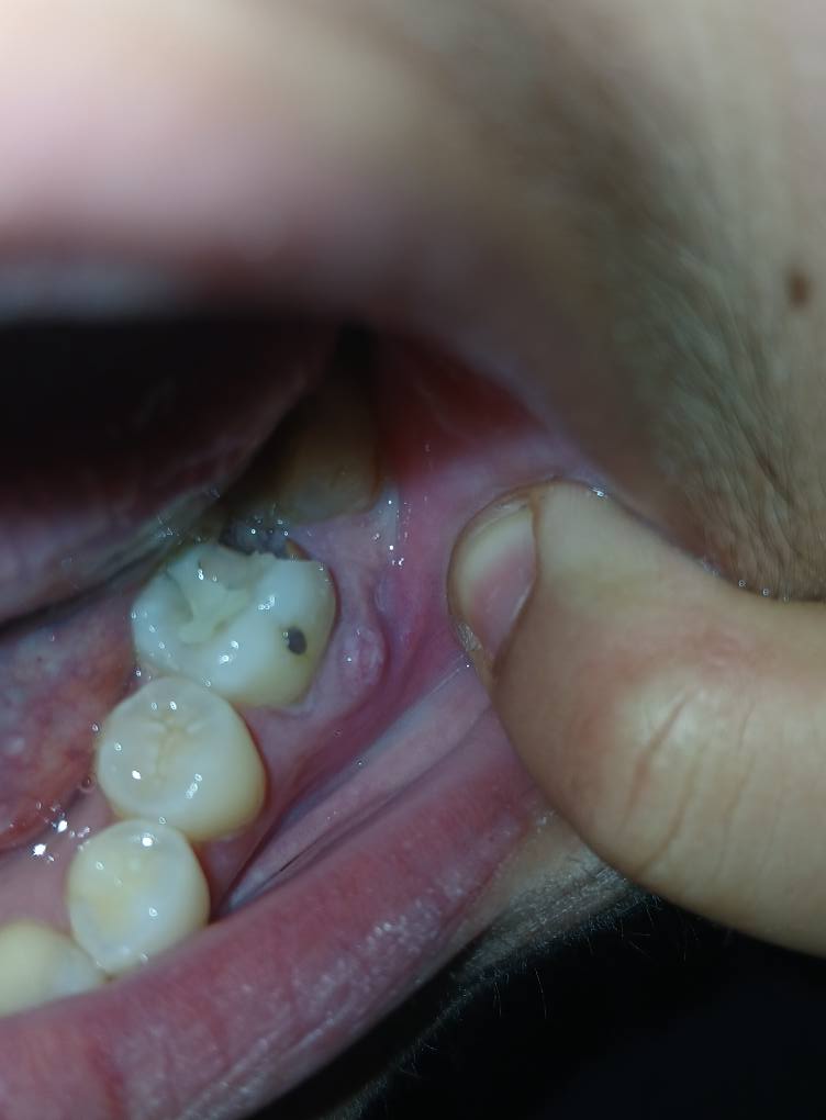
  • Gross Caries (Severe Decay): The lower molar (bottom right of the photo) shows a large, dark cavitation covering most of the occlusal (biting) surface. This indicates extensive tooth structure loss.

  • Likely Pulp Exposure: Given the depth of the dark area on the lower molar, the decay has likely reached the pulp (nerve) of the tooth.

Severe Upper Tooth Decay - Advanced Dental Caries Case

Severity: 

Severe Upper Tooth Decay With Multiple Cavities

Case Overview

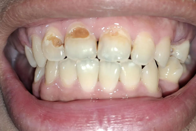
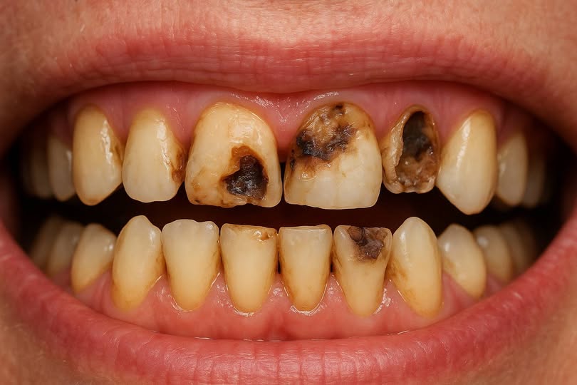
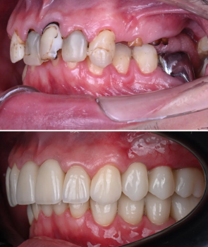
This case shows advanced dental decay affecting multiple upper teeth. Several teeth present with large cavities, enamel breakdown, discoloration, and structural loss. The condition appears long-standing and untreated.

The lower teeth show mild plaque accumulation with possible early decay. The primary concern is severe destruction of the upper teeth with a high risk of infection.


Scale All Teeth

Upper Teeth

  • Multiple visible cavitations

Severe Broken and Infected Teeth Case Analysis With Low-Cost Treatment Options

Severity: 

Teeth Case – Full Analysis and Provisional Diagnosis (Zoom 100%)

Medical disclaimer:
This assessment is based on a single image and patient statement. A confirmed diagnosis requires an in-person dental examination, radiographs, and professional evaluation. Dental infections can become serious and should not be ignored.


1. Visual Findings (Deep Examination)

  • Severely broken teeth:

    • One or more teeth appear fractured down to the root level.

Child Front Teeth Decay Case Analysis and Treatment Timeline

Severity: 

Teeth Case – Full Analysis and Provisional Diagnosis (Zoom 100%)

This evaluation is based only on the provided image. A definitive diagnosis requires a clinical dental examination, appropriate radiographs, and professional assessment by a licensed dentist, particularly for pediatric patients.


1. Visual Findings (Deep Image Review)

  • Upper and lower anterior teeth (front teeth):

Severe Lower Molar Decay: Urgent Restoration and Gum Care Needed

Severity: 

Severe Lower Molar Decay: Urgent Restoration and Gum Care Needed

Case Type: Advanced Dental Caries and Calculus Accumulation


Full Analysis and Diagnosis

Upon examination, the posterior molars exhibit:

  • Extensive caries (tooth decay) on both left and right sides.

  • Possible pulp involvement — dark discoloration and cavity penetration indicate the decay may have reached the pulp chamber.

Failing Molar Fillings with Recurrent Decay – Clinical Case Analysis

Severity: 

Teeth Case Analysis (Based on Photo)

Visual Findings (Zoom 100%)

From the provided image, the lower posterior teeth show:

1. Multiple failing restorations

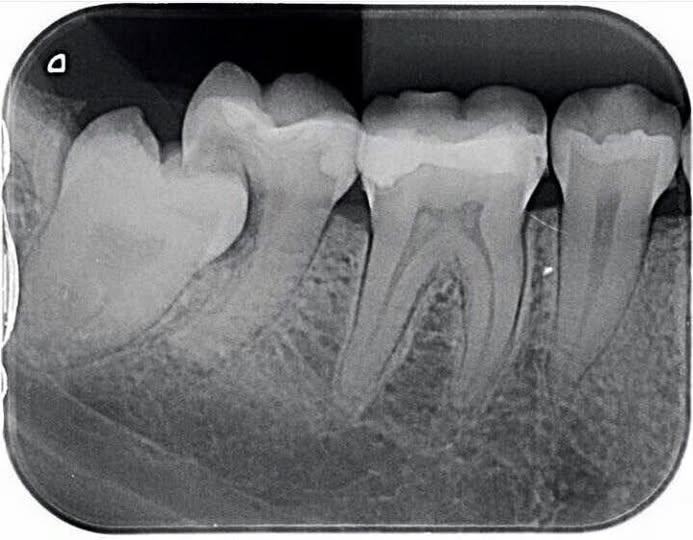
  • Old amalgam fillings with breakdown and marginal leakage

  • Visible dark gaps suggesting secondary caries

2. Deep cavitated lesion

  • The middle molar shows a large open cavity with food debris

Lower Teeth with Amalgam Fillings Case Analysis
Severity: 

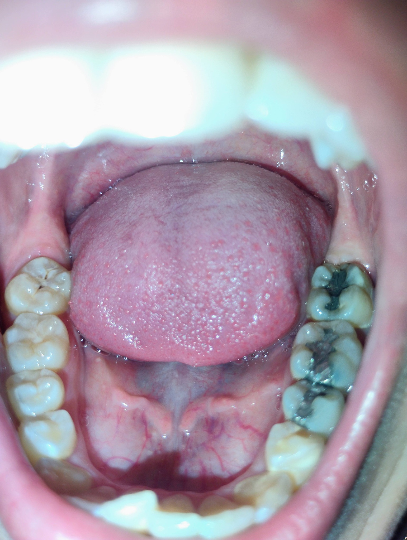
Dental Case Analysis: Lower Teeth with Metallic Fillings and Plaque Accumulation

Case Overview

The image shows the lower dental arch with visible metallic (amalgam) restorations on posterior molars. There are signs of plaque accumulation and dark staining within the grooves of treated teeth.


Zoom 100% Visual Examination

Observed Findings

  • Multiple amalgam (silver) fillings on lower molars

  • Dark discoloration around restoration margins
Severe Tooth Decay and Gum Infection Case Analysis

Severity: 

Dental Case Analysis: Multiple Advanced Tooth Decay With Gum Inflammation

Case Overview (100% Zoom Visual Inspection)

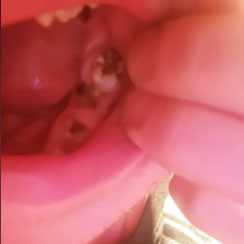
The image shows the upper and lower dental arches with multiple teeth affected by advanced decay, visible dark cavities, broken tooth structure, and inflamed gum tissue. Several teeth show severe enamel and dentin destruction, indicating a high-risk oral condition requiring urgent dental care.

Pages