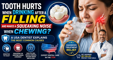
Tooth Hurts When Drinking After a Filling and Makes a Squeaking Noise When Chewing? A USA Dentist Explains the Most Common Causes
Topics:
By a USA Dental Clinical Review Team
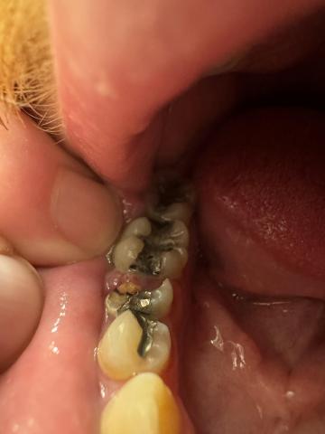
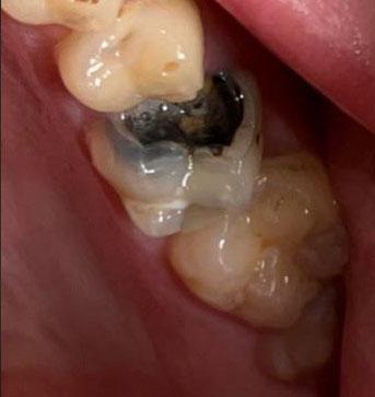
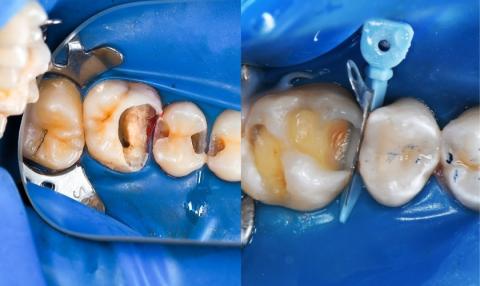
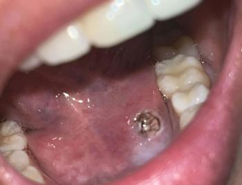
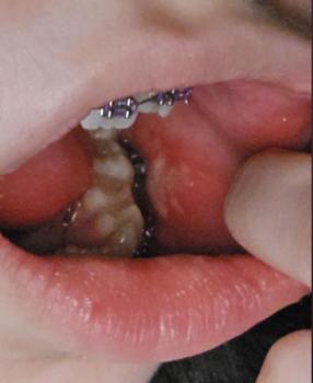
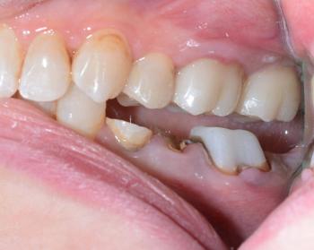
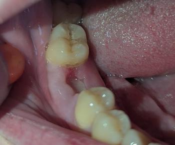
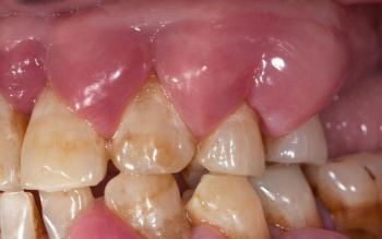
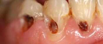
Receiving a new dental filling should relieve pain—not create new symptoms. However, some patients in the United States notice that shortly after a filling, their tooth becomes sensitive when drinking cold or hot beverages and may even produce a squeaking, grating, or rubbing sensation while chewing.
Although these symptoms can be concerning, they are usually explainable and, in many cases, easily corrected by your dentist.