Gum infection

Worsening Gum Swelling and White Spots: Causes, Warning Signs & When to See a Dentist
Severity: 

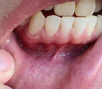
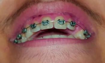
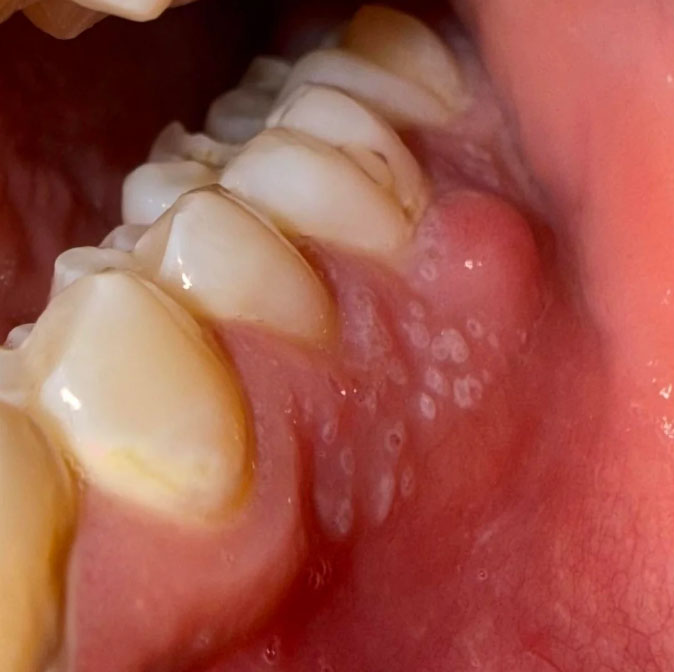
I examined the image at 100% and enlarged it. This is not enough to make a definitive diagnosis, because an intraoral photo cannot show the tooth roots, bone, periodontal pockets, or pulp. But there are several findings that deserve attention—especially because you report that it is getting worse every day.

What I can see

1. Gum/soft-tissue inflammation
Is This Gum Recession? Braces, Gum Inflammation & What Happens in 14 Days

Severity: 

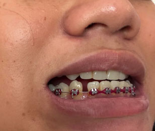
I examined the uploaded photo at 100% and enlarged the lower teeth/gum area. This is a visual assessment only—not a definitive dental diagnosis. A dentist/orthodontist needs periodontal probing and, if indicated, X-rays to determine whether there is true gum recession or bone loss. The American Association of Orthodontists specifically notes that gum health cannot be accurately evaluated from appearance alone.

1. What I can see in this case

Lower front teeth with fixed braces:

Dental X-Ray Case Analysis: Severe Tooth Crowding, Eruption Problems, and What Happens If Treatment Is Delayed 14 Days

Severity: 

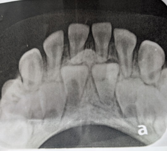
The image appears to be an anterior dental occlusal radiograph, likely showing a developing/mixed-dentition situation. However, an X-ray alone cannot provide a definitive diagnosis or replace an in-person examination.

1. What I can see

The most important finding is crowding/overlap of the anterior teeth with developing or unerupted teeth positioned behind/among the erupted teeth.

Lower Teeth Calculus and Floor-of-Mouth Lesion: Comprehensive Dental Case Analysis and 14-Day Treatment Guide

Severity: 
Teeth Problems: 

Full Dental Case Analysis

1. Overall Photographic Assessment

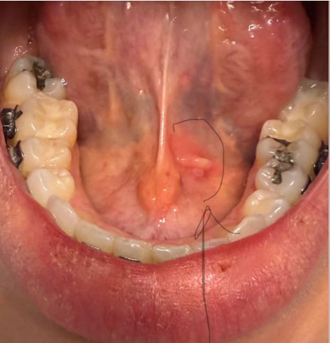
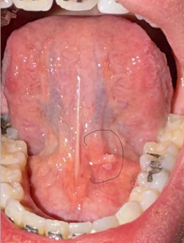
The photograph provides a good view of the lower teeth, tongue, floor of the mouth, and surrounding oral tissues.

The most noticeable dental finding is a visible accumulation of yellowish to brownish material along the tongue-facing surfaces of the lower anterior teeth. This appearance is compatible with dental calculus, commonly called tartar.

There also appears to be localized gingival irritation around the lower teeth.

Dental Case Analysis: Heavy Lower-Tooth Calculus, Possible Gingival Disease, and a Floor-of-Mouth Lesion

Severity: 

This is a photographic assessment, not a definitive diagnosis. The image provides useful visual information, but a dentist must perform an intraoral examination, periodontal probing, and appropriate radiographs before determining the actual diagnosis and severity.

1. Image Examination at 100% Scale

The photograph provides a clear view of the underside of the tongue, the lower anterior teeth, lower posterior teeth, and surrounding soft tissues.

Pages