Chipped Front Teeth Case Analysis and Treatment Options
Severity:
Teeth Problems:
Dental Case Analysis: Chipped and Uneven Front Teeth
Case Overview
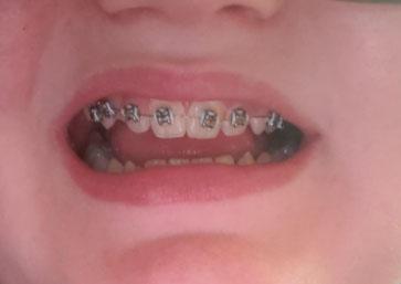
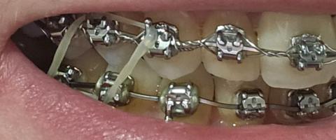
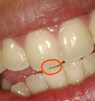
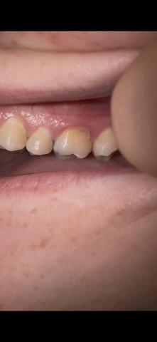
The image shows a side view of the anterior teeth with visible enamel chipping and uneven incisal edges. The teeth appear slightly misaligned with minor surface discoloration.
Zoom 100% Visual Examination
Observed Findings
-
Small to moderate enamel chips on front teeth
-
Uneven incisal (biting) edges
-
Mild yellow discoloration
-
Slight spacing irregularity