Mild

Urgent Dental Bonding for Chipped Front Tooth with Mini Fracture,

Severity: 

Initial Image Analysis

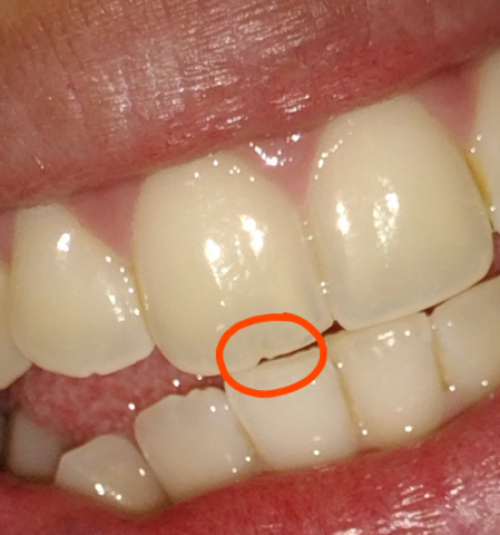
  • Image Observations: The picture shows the front teeth (incisors). The central incisor (the main front tooth) on the viewer's left has a chipped or fractured corner (incisal angle). A thin, dark, or gray line—the "mini fracture"—appears to run vertically or slightly obliquely from the chipped edge toward the gum line.

  • Client Statement: "I have chipped front tooth that has mini fracture in it, should I bond it or smooth it out?"


Emergency Dental Appointment for Failed Crown or Deep Cavity

Severity: 

Initial Image Analysis

  • Image Observations: The picture shows the lower right side of the mouth (Mandibular quadrant).

    • The tooth farthest back (most posterior) appears to have a restoration (filling or inlay/onlay) or possibly a crown which exhibits a large, dark, and deep-looking defect or void on its biting surface (occlusal surface). The defect appears dark brown/black with a distinct border.

Finding a Dental Clinic for Examination of Dark Tooth Spots

Severity: 

Initial Image Analysis

  • Image Observations: The picture shows the front teeth with small, dark, punctate (dot-like) spots or specks, primarily located close to the gum line (gingival margin). The spots are circled in red for emphasis. The surrounding gum tissue (gingiva) appears generally pink and healthy in the visible area.

  • Client Statement: "Had it some days now. Can't remove with brush."


Lower anterior cervical roughness - likely calculus or non-carious wear; cleaning and possible restoration recommended

Severity: 

Quick clinical impression

  • The lower anterior teeth show rough, yellowish/cream areas at the cervical (gumline) region of several incisors.

  • Most likely causes (photo only — no probing/X-ray): tartar/calculus buildup with superficial stain, and/or non-carious cervical wear (abrasion/erosion) exposing dentine. The rough texture could be hardened calculus or eroded tooth surface.

Occlusal fracture / caries - urgent assessment and temporary protection recommended

Severity: 
Teeth Problems: 

Quick clinical impression

  • The picture shows occlusal breakdown / possible caries or fractured cusp on the last visible molar (upper right in the photo). The tooth surface looks cavitated and stained; one area looks like loss of enamel/restoration.

  • I don’t see obvious swelling or pus in the photo, and I can’t test percussion/mobility or pulp vitality from a photo — those are needed for definitive diagnosis.

Pages