Moderate

Severe Tartar on Lower Front Teeth: Hidden Gum Disease, Bone Loss Risk, Treatment Process & 14-Day Healing Timeline

Severity: 
Teeth Problems: 

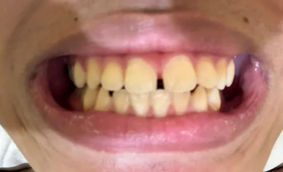
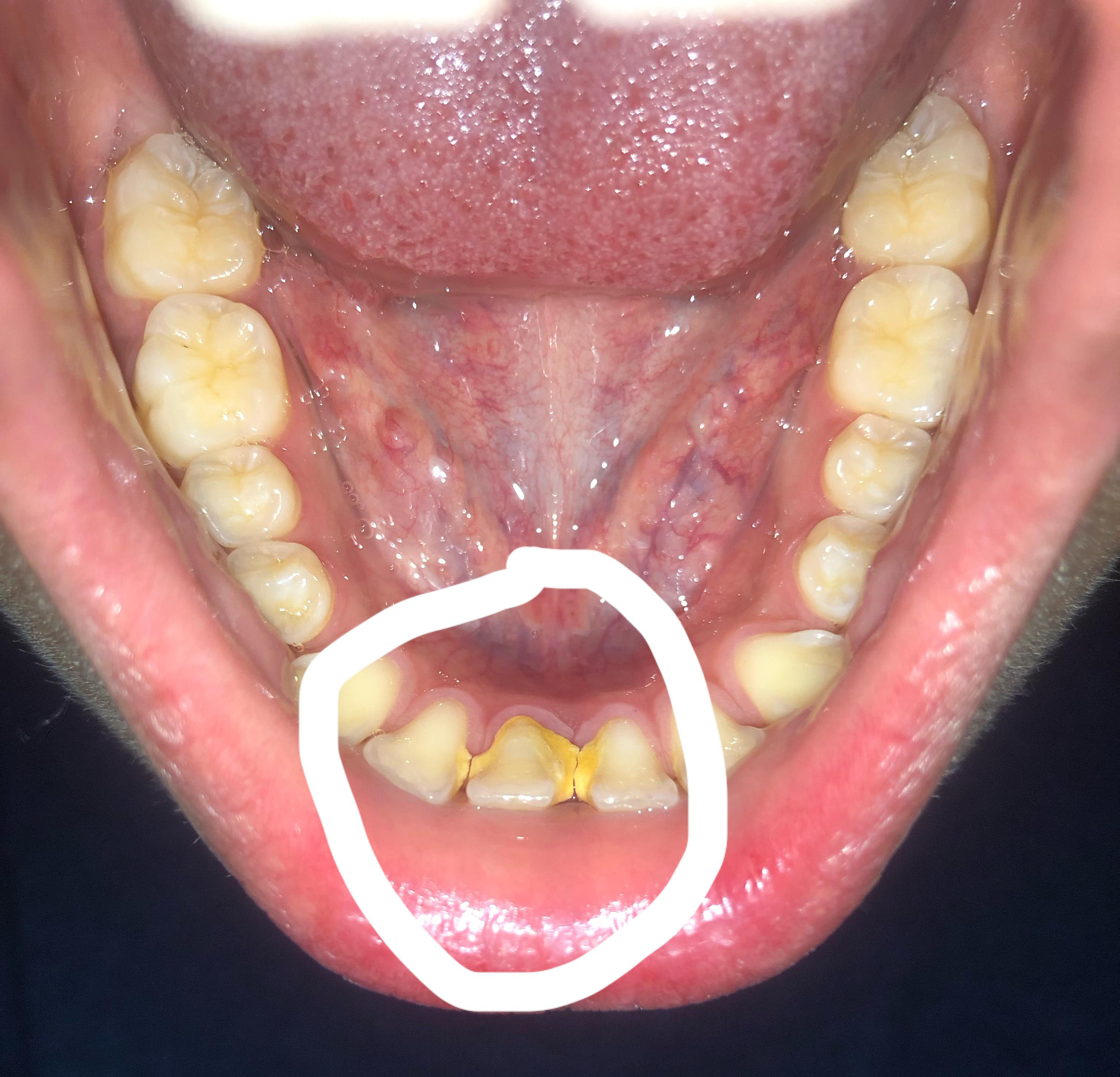
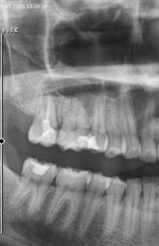
I examined the photograph at 100% resolution and focused especially on the circled lower-front region. This is a clinical-photo assessment, not a definitive diagnosis; periodontal probing and appropriate X-rays are needed to determine whether there is bone or attachment loss.

Severe Tartar Buildup on Lower Front Teeth: Gum Inflammation, Treatment, 14-Day Healing Timeline & Risk of Progression

Severity: 

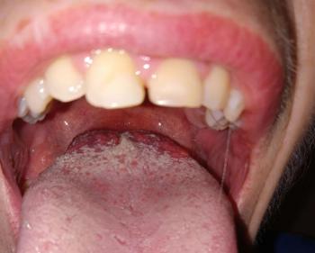
This is a clinical photo rather than an X-ray, so I can assess visible plaque/calculus, gum appearance, tooth alignment, and staining, but I cannot determine bone loss or periodontal pocket depth from the photo alone.

Main finding

The most obvious issue is substantial calculus (tartar) accumulation on the lower front teeth, particularly around the lingual/cervical surfaces. There is also visible gingival redness/inflammation.

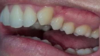
The lower incisors appear crowded/rotated, which can make plaque and calculus much easier to accumulate.

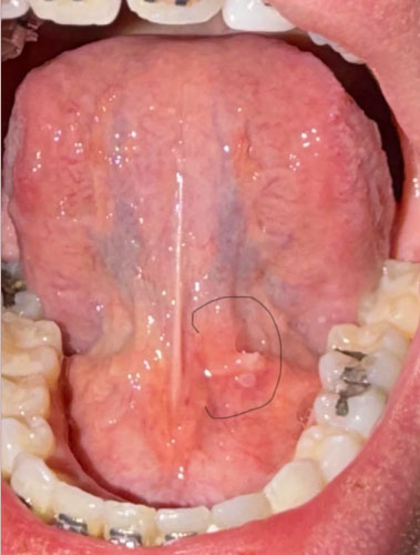
Lower Teeth Calculus and Floor-of-Mouth Lesion: Comprehensive Dental Case Analysis and 14-Day Treatment Guide

Severity: 
Teeth Problems: 

Full Dental Case Analysis

1. Overall Photographic Assessment

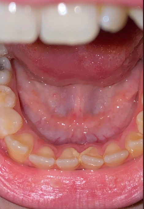
The photograph provides a good view of the lower teeth, tongue, floor of the mouth, and surrounding oral tissues.

The most noticeable dental finding is a visible accumulation of yellowish to brownish material along the tongue-facing surfaces of the lower anterior teeth. This appearance is compatible with dental calculus, commonly called tartar.

There also appears to be localized gingival irritation around the lower teeth.

Dental Case Analysis: Heavy Lower-Tooth Calculus, Possible Gingival Disease, and a Floor-of-Mouth Lesion

Severity: 

This is a photographic assessment, not a definitive diagnosis. The image provides useful visual information, but a dentist must perform an intraoral examination, periodontal probing, and appropriate radiographs before determining the actual diagnosis and severity.

1. Image Examination at 100% Scale

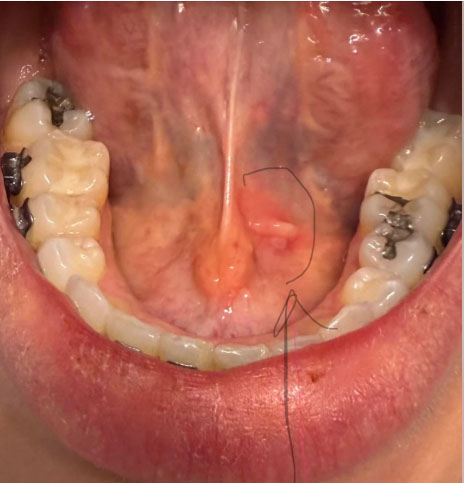
The photograph provides a clear view of the underside of the tongue, the lower anterior teeth, lower posterior teeth, and surrounding soft tissues.

Heavy Dental Calculus and Gingival Inflammation: Comprehensive Clinical Assessment, Treatment Timeline, and 14-Day Healing Guide

Severity: 

A dentist would need periodontal probing, bite/occlusal examination, and usually appropriate radiographs to determine bone loss and whether there is an underlying dental infection.

What I can see at 100% view

The most obvious finding is substantial hardened calculus/tartar on the tongue-side (lingual) surfaces of the lower front teeth, particularly around the lower incisors. There also appears to be localized redness/inflammation of the soft tissue under the tongue, near the floor-of-mouth/frenulum region.

Pages