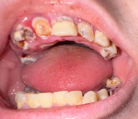
Severe Rooted Tooth Decay and Infection Treatment
Severity:
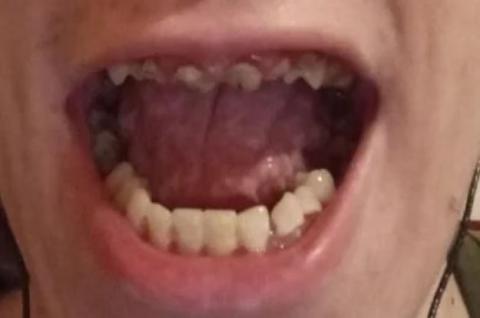
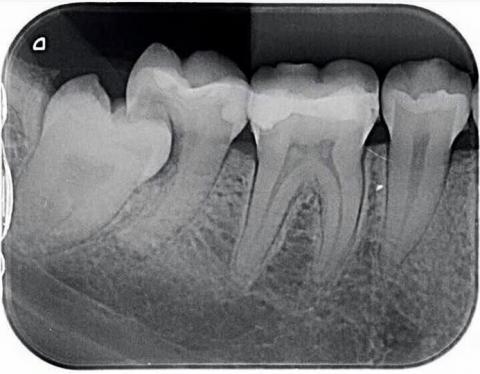
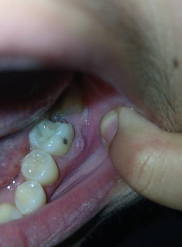
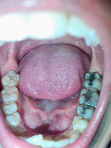
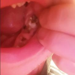
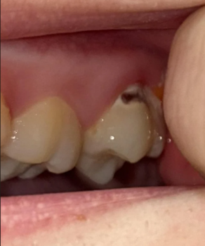
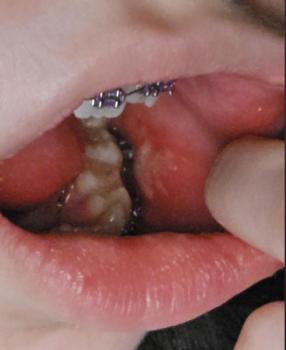
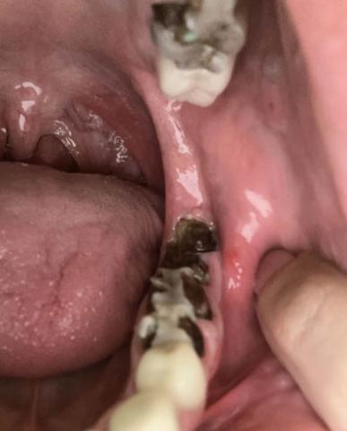
This case shows severe tooth decay affecting the back teeth, with decay extending close to or into the tooth roots. Dark, broken tooth structure is visible, which indicates long-standing dental caries. The surrounding gum tissue appears irritated, increasing the risk of infection.
Rooted tooth decay usually develops when cavities are left untreated for a long time. Bacteria spread deeper into the tooth and reach the root area, making the tooth weak, painful, and difficult to save.