Severe Upper Tooth Decay - Advanced Dental Caries Case
Severity:
Teeth Problems:
Severe Upper Tooth Decay With Multiple Cavities
Case Overview
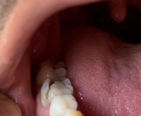
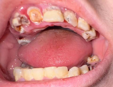
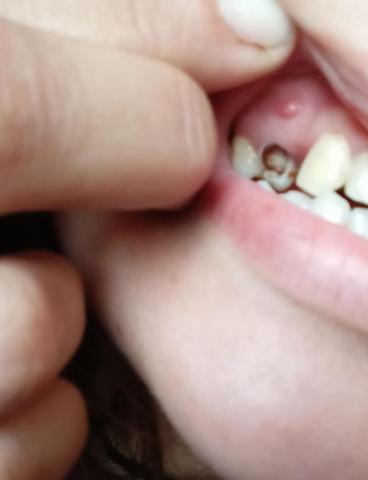
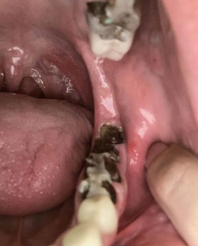
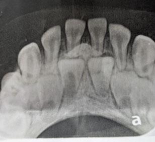
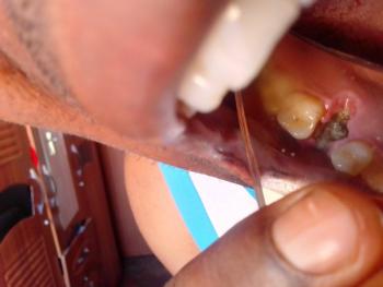
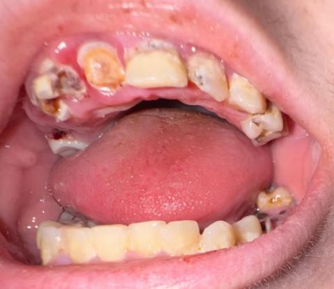
This case shows advanced dental decay affecting multiple upper teeth. Several teeth present with large cavities, enamel breakdown, discoloration, and structural loss. The condition appears long-standing and untreated.
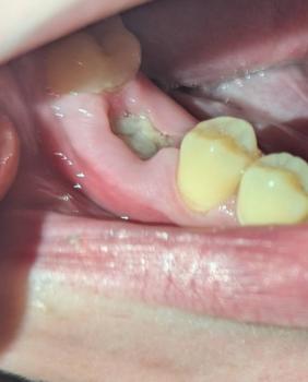
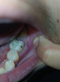
The lower teeth show mild plaque accumulation with possible early decay. The primary concern is severe destruction of the upper teeth with a high risk of infection.
Scale All Teeth
Upper Teeth
-
Multiple visible cavitations