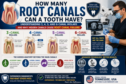
Root canal infection

Post Root Canal Infection With Sutures Case Analysis and Treatment Guide

Severity: 

What Is Seen in This Case

The image shows a treated tooth with sutures placed on the surrounding gum tissue. The area appears reddened and swollen, indicating recent root canal–related surgical treatment or management of an infection at the root area. The presence of sutures suggests that the infection required surgical access, drainage, or gum repair.

This is a normal appearance shortly after treatment but requires close monitoring.

Severe Front Tooth Decay Near the Gum Line | Can a Root Canal Save the Tooth

Severity: 

Severe Cervical Decay on Upper Front Tooth – Full Dental Case Analysis (100% Zoom)

Medical Disclaimer

This is an educational, image-based analysis only. A definitive diagnosis requires an in-person dental examination, X-rays, and vitality testing. The findings below are based on visible clinical signs and standard dental assessment protocols.

Severe Tooth Decay and Multiple Cavities | Advanced Dental Case Analysis

Severity: 

Severe Multi-Tooth Decay With Advanced Cavities – Full Dental Case Analysis (100% Zoom)

Medical Disclaimer
This assessment is image-based and for educational purposes only. A confirmed diagnosis requires an in-clinic dental examination, X-rays, and vitality testing. However, the visible findings indicate a serious and urgent dental condition that should not be delayed.

Exposed Dental Pulp and Infection | Root Canal Case Analysis and Treatment Guide

Severity: 

Exposed Pulp With Advanced Infection – Root Canal–Level Dental Case (100% Zoom Analysis)

Clinical Disclaimer
This is an image-based educational analysis. A definitive diagnosis requires an in-clinic examination, vitality testing, and dental X-rays. The visual evidence, however, indicates a severe and urgent condition requiring professional treatment.

Severe Root Caries Near Gum Line | Dental Case Analysis and Treatment Timeline

Severity: 

Severe Cervical & Root Caries – Full Clinical Analysis (Zoomed View)

Case Overview (100% Zoom Interpretation)

The image shows advanced dental caries located at the cervical and root surfaces of multiple adjacent teeth, very close to the gum line. There is visible cavitation, darkened dentin, and signs consistent with gingival recession, exposing the root surfaces. This pattern suggests an active, progressing disease process, not a superficial stain.

Pages