Dry Socket vs Infection in California USA: Complete 2026 Dental Expert Guide
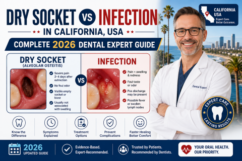
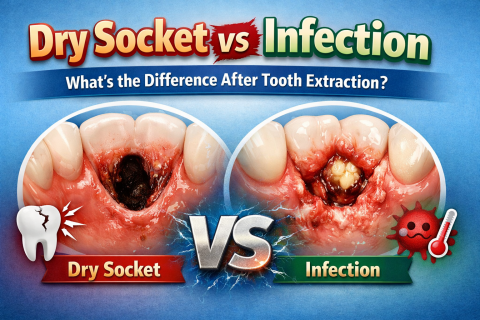
Tooth extraction and dental implant procedures are among the most common oral surgeries performed across California, USA. While most patients heal normally, some experience painful complications during recovery. Two of the most misunderstood problems are dry socket and dental infection.
Many patients confuse the two because both conditions may occur after tooth extraction, wisdom tooth removal, or dental implant surgery. However, they are very different problems requiring different treatment approaches.