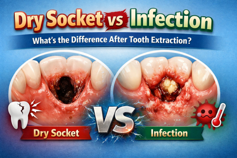
Dry Socket vs Infection: What’s the Difference After Tooth Extraction?
After a tooth extraction, many patients ask:
“Is this dry socket or infection?”
“Why is my pain getting worse?”
For busy workers, the common mistake is waiting.
They continue working long shifts.
They only file a long day off when the pain becomes unbearable.
By then, treatment becomes more complicated — and more expensive.
Understanding the difference early can save you time, money, and stress.
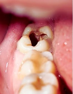
What Is Dry Socket?
Dry socket (alveolar osteitis) happens when the protective blood clot falls out or dissolves too early.