Dental Case: Problems

Why Do My Front Teeth Hurt? Decay Case Analysis and Treatment Guide

Severity: 

Dental Case Analysis: Front Teeth Decay with Active Pain Complaint

Client Concern

Client said:
“Hi, my teeth hurt. Any observations from these please?”


Case Overview

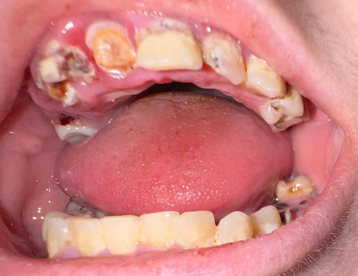
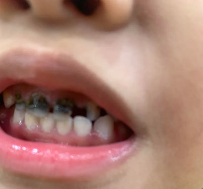
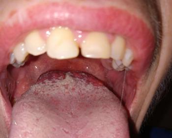
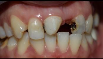
The image shows the upper and lower front teeth with visible brown enamel defects and decay on the upper incisors. There is also visible misalignment and plaque accumulation.

Pain reported by the client is consistent with the visual findings.

Severe Tooth Decay With Gum Infection - Dental Case Analysis

Severity: 

Dental Case Analysis – Severe Tooth Decay With Localized Gum Infection

Case Overview

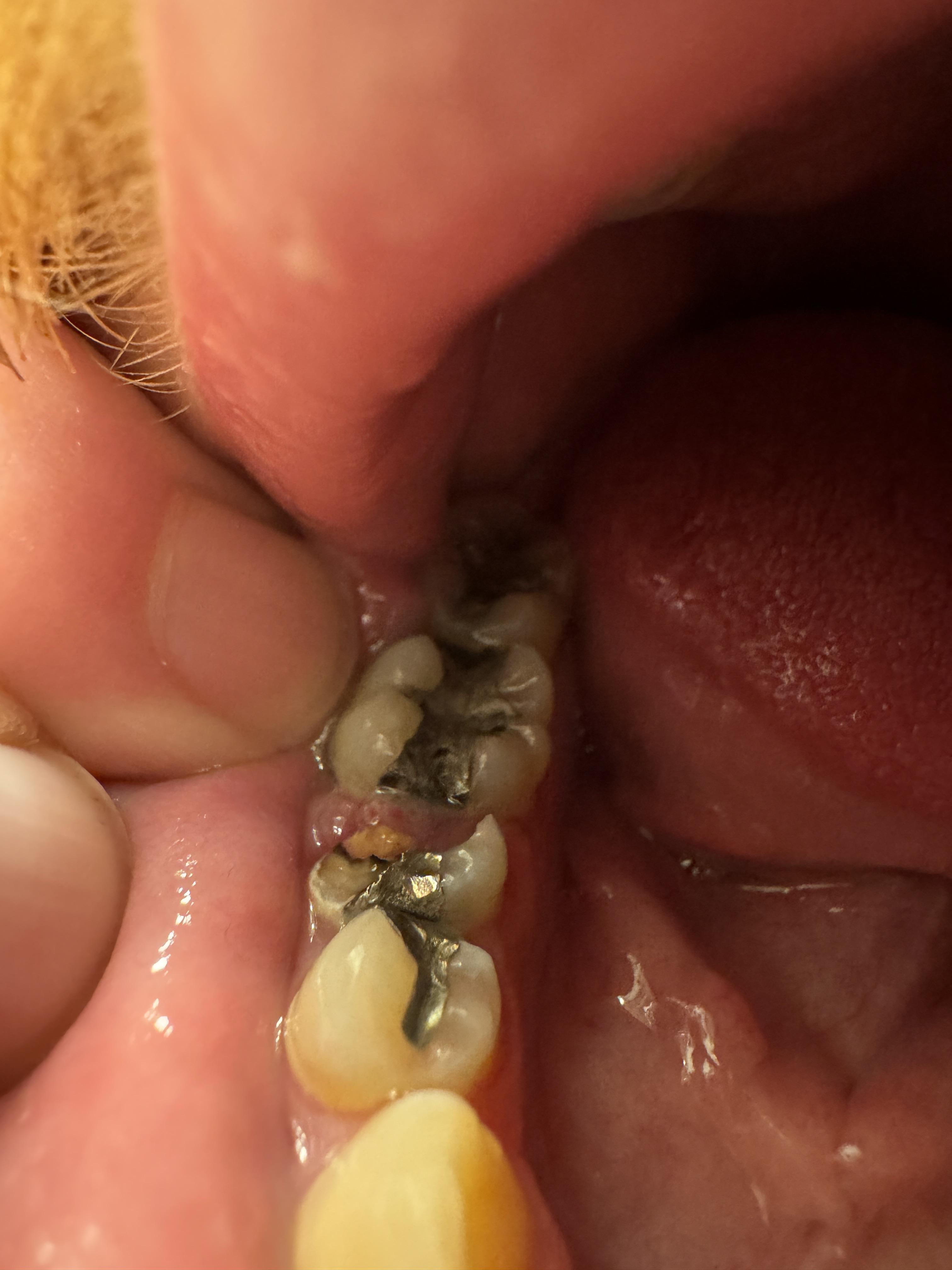
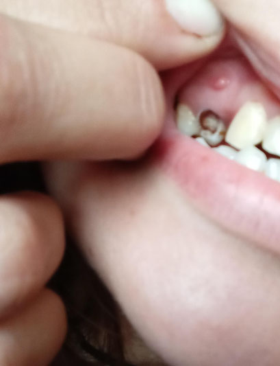
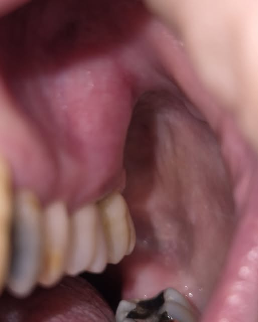
This intra-oral image shows a tooth with advanced decay and an associated localized gum swelling above the affected tooth. The appearance suggests an active dental infection likely originating from deep caries that has reached the pulp and spread toward the surrounding soft tissue.


Full Analysis and Diagnosis

Clinical Findings

  • Deep cavity with visible tooth structure breakdown

Failing Molar Fillings with Recurrent Decay – Clinical Case Analysis

Severity: 

Teeth Case Analysis (Based on Photo)

Visual Findings (Zoom 100%)

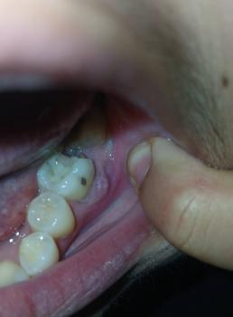
From the provided image, the lower posterior teeth show:

1. Multiple failing restorations

  • Old amalgam fillings with breakdown and marginal leakage

  • Visible dark gaps suggesting secondary caries

2. Deep cavitated lesion

  • The middle molar shows a large open cavity with food debris

Severe Rooted Tooth Decay and Infection Treatment

Severity: 

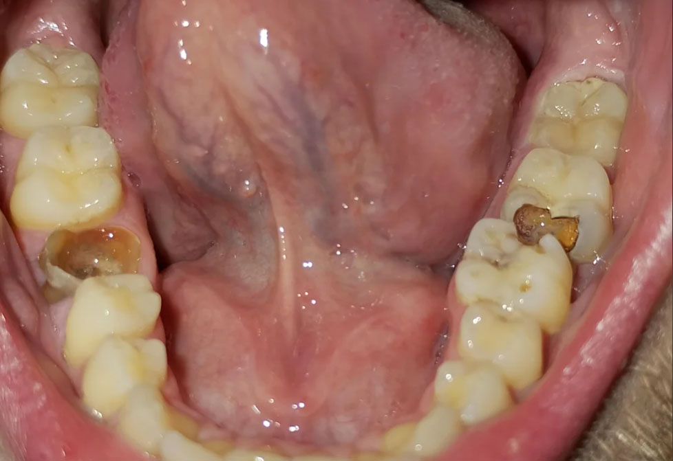
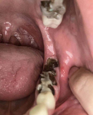
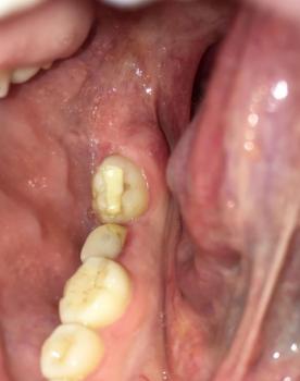
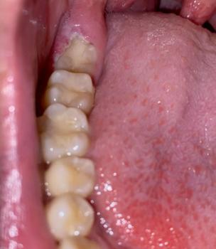
This case shows severe tooth decay affecting the back teeth, with decay extending close to or into the tooth roots. Dark, broken tooth structure is visible, which indicates long-standing dental caries. The surrounding gum tissue appears irritated, increasing the risk of infection.

Rooted tooth decay usually develops when cavities are left untreated for a long time. Bacteria spread deeper into the tooth and reach the root area, making the tooth weak, painful, and difficult to save.

Severe Tooth Decay Treatment in the Philippines: Treatment Options, Costs, and Before & After Results

Severity: 
Teeth Problems: 

Case Analysis: Advanced Dental Caries and Enamel Erosion

Clinical Observation

Upon visual examination of the image:

Lower Teeth with Amalgam Fillings Case Analysis

Severity: 

Dental Case Analysis: Lower Teeth with Metallic Fillings and Plaque Accumulation

Case Overview

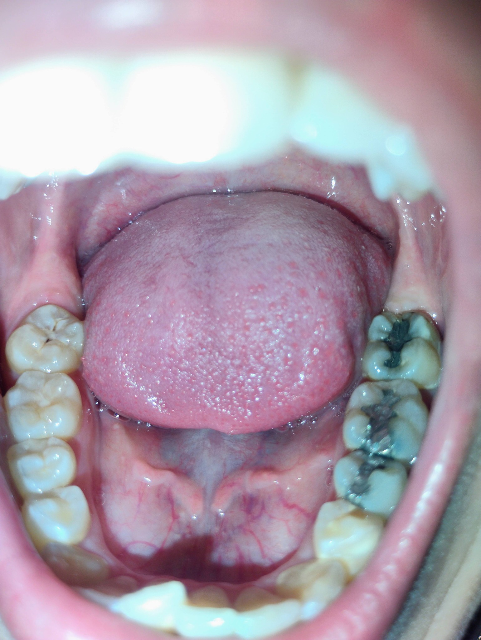
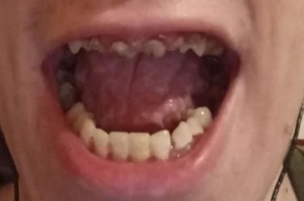
The image shows the lower dental arch with visible metallic (amalgam) restorations on posterior molars. There are signs of plaque accumulation and dark staining within the grooves of treated teeth.


Zoom 100% Visual Examination

Observed Findings

  • Multiple amalgam (silver) fillings on lower molars

  • Dark discoloration around restoration margins

Tooth Decay and Gum Infection: Causes, Treatment, and Healing in 7 Days

Severity: 
Teeth Problems: 

Case Analysis: Tooth Decay and Gum Inflammation – Early Warning of Dental Infection

Client’s Complaint

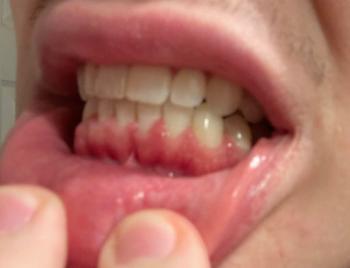
“My back teeth hurt and smell bad. I noticed dark spots and some swelling around the gum area.”


Clinical Observation (From Image)

After close examination of the photo:

  • Visible dark spot (cavity) on one molar — likely tooth decay or caries.

Severe Night Toothache & Deep Cavity Treatment in California, US

Severity: 

The image strongly suggests a case of Severe Tooth Decay and Possible Pulp Exposure. The pain shown in the right panel indicates a severe toothache, which often means the decay has progressed to the innermost part of the tooth (the pulp), causing irreversible pulpitis or a dental abscess. This requires urgent professional attention.

Do not attempt to self-treat this condition. Use the provided directory link to find a nearby clinic immediately.


The Hidden Threat: What Back Tooth Pain and Bleeding Gums Could Mean for Your Health

Severity: 

Deep Examination & Analysis

Based on the image provided, there are several significant dental health concerns that require immediate professional attention:

  • Gross Caries (Severe Decay): The lower molar (bottom right of the photo) shows a large, dark cavitation covering most of the occlusal (biting) surface. This indicates extensive tooth structure loss.

  • Likely Pulp Exposure: Given the depth of the dark area on the lower molar, the decay has likely reached the pulp (nerve) of the tooth.

Pages