Full Mouth Dental Rehabilitation Case – Before & After Analysis (14-Day Implant Possibility)
Severity:
Teeth Problems:
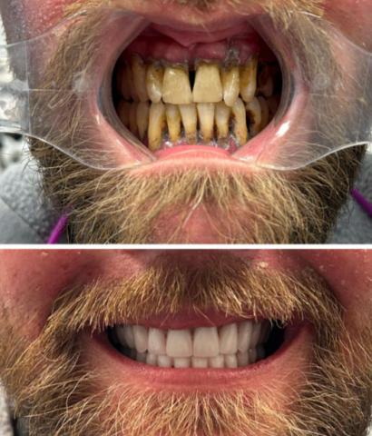
Teeth Case Analysis (Before & After)
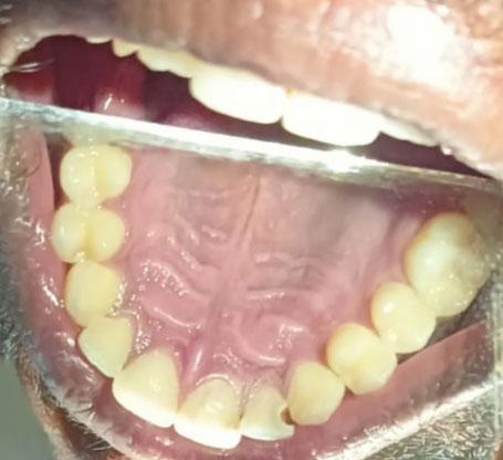
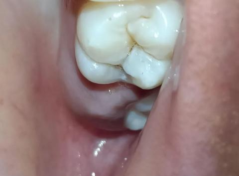
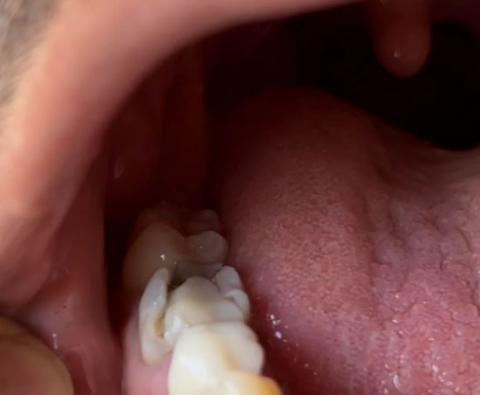
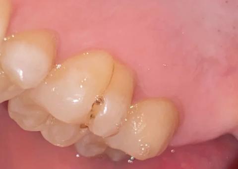
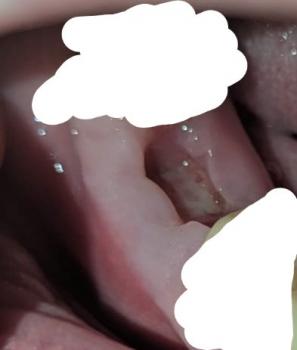
Clinical Observation (Zoom-Level Review)
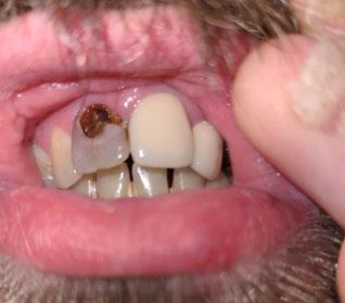
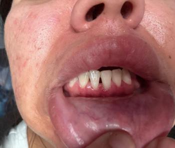
From the image provided:
Before Condition
-
Severe plaque and calculus buildup (heavy tartar deposits)
-
Advanced discoloration (brown/yellow staining)
-
Gingival inflammation (red, swollen gums)
-
Possible periodontal disease (bone loss likely)
-
Multiple cervical caries (dark lesions near gumline)
-
Tooth wear and uneven edges