Dental Abscess Still Swollen After Antibiotics? Expert Dentist Explains When Drainage Is Needed
Topics:
Real Dental Abscess Case: Swollen Gum With Minimal Improvement After 3 Days
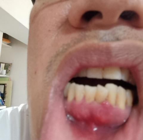
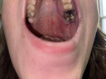
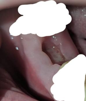
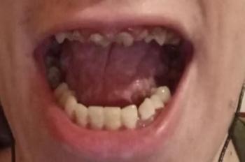
Karl noticed something unusual developing in his lower gum area. The swelling became red, enlarged, and painful. The gum looked like it was “swallowing” part of the tooth area, a common description patients use when pressure builds from a dental abscess.
He immediately visited a dental clinic.
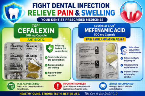
The dental assistant advised him to take: