Transforming Smiles, Restoring Confidence.
Bad Smell from Gums After Tooth Removal? Expert Dentist Explains Causes and Fixes
Image:
Severity:
Teeth Problems:
Case summary
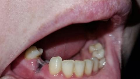
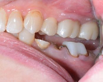
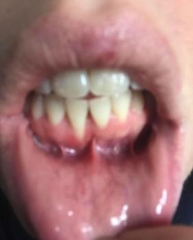
Based on the photos (zoomed and reviewed from multiple angles): there is a partially healed/missing-molar area in the lower jaw with a thin, whitish/gray string-like band or tissue running between the back area and the adjacent teeth. The adjacent gums look slightly inflamed. The patient reports pain and bad smell. This most commonly represents a localized infection or retained foreign material (food, suture/retraction cord fragment, bone sequestrum) with secondary gum inflammation and possible sinus tract formation.
Key findings (from the images)
-
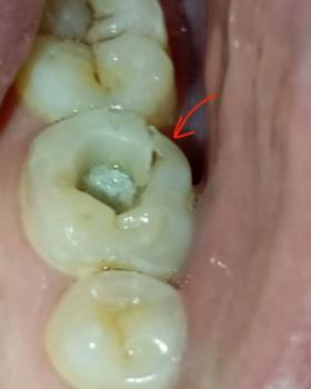
Missing or heavily restored posterior molar region visible.
-
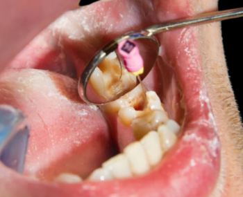
A pale/gray fibrous strand or material bridging the extraction/defect site to the adjacent tooth.
-
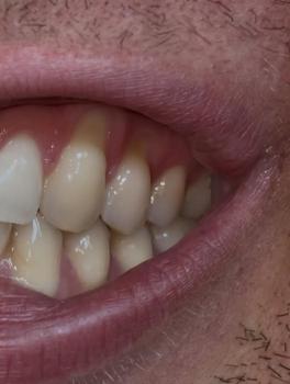
Localized gingival redness and mild swelling.
-
Evidence of plaque/debris on adjacent teeth that could feed infection.
-
Malodor reported by patient — suggests bacterial activity/decay or draining infection.
Differential diagnosis (most likely → less likely)
-
Localized draining sinus / fistula from a root infection — chronic infection trying to drain through soft tissue.
-
Retained foreign material (food plug / retraction cord / suture) causing persistent irritation and infection.
-
Bone sequestrum (small dead bone fragment) protruding under the gum and irritating tissue.
-
Granulation tissue or epithelialized sinus tract (chronic healing tissue that can look like a strip).
-
Periodontal abscess or chronic gingival infection at the site with food entrapment.
Best method for fastest, reliable recovery (summary)
Definitive control of the source of infection is fastest: urgent clinical exam → targeted X-ray (periapical/CBCT if available) → remove the offending material (debridement / removal of bone fragment / curettage) and either treat the source tooth (root canal) or extract if non-restorable. Drainage + local irrigation + appropriate antibiotics (only when indicated) + good oral hygiene speeds recovery.
Step-by-step recommended process (what the dentist will do)
-
Urgent appointment and X-ray (periapical view; CBCT if available) to identify source (root tip, sequestrum, retained fragment).
-
Clinical inspection under local anaesthesia to probe the area and determine if the strand is foreign material, granulation tissue, or bone.
-
Remove foreign material / curettage the socket (clean and irrigate thoroughly). If a bone sequestrum is present it will be removed.
-
Drainage if there is pus; collect sample/culture if chronic or recurrent.
-
Definitive tooth treatment: either root canal retreatment (if tooth salvageable) or extraction (if tooth is hopeless).
-
Antibiotics only if there are systemic signs or spreading infection (dentist will prescribe appropriate drug and dose).
-
Suture or allow secondary intention healing depending on wound; post-op instructions.
-
Follow-up at 1 week and again at 2–4 weeks to confirm healing; further restorative work thereafter (crown, bridge, implant) if needed.
Immediate home care until you can see a dentist
-
Do not pick, cut, or try to pull the strand yourself. That can push infection deeper.
-
Rinse gently with warm salt water (1 tsp salt in 250 mL warm water) 3–4 times daily.
-
Use a 0.12% chlorhexidine rinse if available and advised by a clinician.
-
Soft diet; avoid chewing on that side.
-
Over-the-counter pain relief (ibuprofen or paracetamol per label/doctor’s advice).
-
Keep good oral hygiene (gentle brushing, interdental cleaning away from the painful area).
Expected healing timeline
-
Symptom relief (after drainage + antibiotics if needed): often within 24–72 hours.
-
Soft tissue healing: 7–14 days for the gum to close and inflammation to subside.
-
Complete bone/socket remodeling (if extraction): weeks to months (initial bone fill by 6–8 weeks, remodeling longer).
-
If root canal is done: pain typically improves in 48–72 hours; full comfort within 1–3 weeks.
What happens if treatment is delayed 30 days
If left untreated for 2–4 weeks the problem commonly worsens:
-
Persistent or increasing pain and bad taste/odor.
-
Development of a frank abscess with pus and facial swelling.
-
Spread of infection into adjacent teeth or jawbone (osteomyelitis) requiring more extensive surgery.
-
Loss of the adjacent tooth or need for extraction rather than conservative root canal therapy.
-
In rare severe cases, spreading cellulitis (medical emergency) — seek immediate care for rapidly spreading swelling, fever, difficulty breathing or swallowing.
Urgent warning signs — go for immediate care or emergency room
-
Rapidly increasing facial swelling, fever, or difficulty breathing/swallowing.
-
Severe uncontrolled pain, spreading redness beyond the mouth.
If any of these occur, seek emergency medical/dental care immediately.
Practical next step and local help
Book an urgent exam with a dentist so they can X-ray and remove the source. Use the clinic directory you provided to find a nearby clinic and book now:
https://cebudentalimplants.com/map-dental-clinic
CASE Comment
This appears to be a localized, treatable dental infection or retained material — but it will not resolve reliably on its own. Early professional cleaning/removal of the source + targeted dental treatment (RCT or extraction) gives the fastest recovery and highest chance to save adjacent teeth. Please arrange a dental visit within 48–72 hours if possible.
If you want, I can draft a short urgent-message template you can copy-paste to a clinic booking form (asking for “urgent exam + X-ray + likely drainage/removal of foreign material”) — tell me which clinic you want to contact and I’ll prepare it.
Teeth Case
 |
 |
 |
 |
 |
 |