Where Bright Smiles Begin.

What Is the Worst-Case Scenario for Dental Bone Grafting? A USA Expert Dentist Explains Risks, Warning Signs, and How to Protect Your Health
Image:
Severity:
Teeth Problems:
Dental bone grafting is one of the most predictable and successful procedures in modern dentistry. Every year, thousands of patients across the United States undergo bone grafting to rebuild lost jawbone, prepare for dental implants, preserve extraction sites, and restore oral function.
For most patients, healing progresses smoothly, and the graft successfully integrates with the surrounding bone. However, it is natural to wonder about potential risks before any surgical procedure. Patients frequently ask me, "What is the worst thing that could happen after a dental bone graft?"
As a dentist, I believe patients deserve honest and balanced information. While severe complications are uncommon, understanding them helps you make informed decisions and recognize warning signs early if problems develop.
The reality is that the vast majority of bone graft complications can be successfully managed when identified quickly. Most serious outcomes occur when infections, healing problems, or underlying health issues are ignored or left untreated.
This article explains the worst-case scenarios associated with dental bone grafting, the factors that increase risk, warning signs to watch for, and practical steps you can take to improve your chances of a successful recovery.
Understanding Dental Bone Grafting
Before discussing complications, it helps to understand what a bone graft actually does.
A dental bone graft is designed to replace missing bone in the jaw. The graft acts as a scaffold that encourages your body to produce new living bone tissue.
Over time, your natural bone gradually replaces or integrates with the graft material.
Bone grafting may be performed for:
- Dental implant preparation
- Socket preservation after tooth extraction
- Ridge augmentation
- Sinus lift procedures
- Repair of bone defects caused by infection
- Trauma reconstruction
- Periodontal disease treatment
When healing occurs normally, the result is a stronger and healthier jawbone capable of supporting future restorations.
How Successful Is Dental Bone Grafting?
Patients are often surprised to learn that dental bone grafting has a very high success rate.
Modern techniques, improved imaging technology, advanced graft materials, and better surgical planning have dramatically improved outcomes.
Most bone grafts heal without major complications when:
- The patient follows instructions carefully
- Oral hygiene is maintained
- Smoking is avoided
- Medical conditions are controlled
- The procedure is performed by an experienced provider
Although success rates are high, no surgical procedure is completely risk-free.
Worst-Case Scenarios After Dental Bone Grafting: Complications, Warning Signs, and Expert Guidance
Understanding the Most Serious Risks of Dental Bone Grafting
Dental bone grafting is a surgical procedure used to rebuild or increase jawbone volume, often to prepare a patient for dental implant placement. Although many patients heal without major complications, the procedure is not risk-free. In some cases, infection, graft exposure, inadequate bone regeneration, or problems involving nearby anatomical structures can affect the final result.
From a dental case-analysis perspective, the most important question is not simply whether a complication can occur. It is whether the patient's symptoms are following an expected healing pattern, whether the graft is integrating successfully, and whether early intervention is needed.
The American Association of Oral and Maxillofacial Surgeons explains that bone graft materials can come from the patient's own body, a donor source, an animal-derived source, or synthetic materials. The choice depends on the defect, the surgical technique, and the treatment plan.
Important clinical principle: A complication does not automatically mean that the graft has failed permanently. Some problems can be treated while preserving part or all of the graft. Other cases require removal of infected material, additional reconstruction, or a revised implant treatment plan.
1. Severe Infection of the Bone Graft
Infection is one of the complications that requires particular attention after oral surgery. It may involve the soft tissue surrounding the graft or, in more serious cases, the grafted bone and adjacent jawbone.
Symptoms that require evaluation
Patients should contact their treating dentist or oral surgeon promptly if they develop:
-
Swelling that continues to increase instead of gradually improving.
-
Pain that becomes more severe after initially improving.
-
Pus or persistent discharge from the surgical site.
-
Fever or chills accompanied by worsening oral symptoms.
-
Increasing redness, tenderness, or an unpleasant taste associated with discharge.
-
Wound breakdown or visible graft material accompanied by worsening symptoms.
Mild swelling, bruising, and discomfort can be expected after surgery. Swelling often peaks around the second or third day, although the course varies according to the procedure. The direction of change is important: progressively worsening symptoms should not be dismissed as normal healing.
How is a suspected infection managed?
A dentist or oral surgeon will assess the surgical site and may obtain appropriate imaging or other investigations. Treatment depends on the severity and extent of the infection and can involve drainage, surgical cleaning, antibiotics when indicated, or removal of compromised graft material.
Antibiotics alone may not resolve an infection if an abscess requires drainage or infected material needs surgical treatment. Patients should not start leftover antibiotics, change prescribed doses, or assume that antibiotics guarantee graft success.
Expert case insight: When pain and swelling worsen rather than improve, clinical examination is more reliable than trying to diagnose graft failure from symptoms alone.
2. Bone Graft Failure or Inadequate Bone Formation
A graft does not always produce enough healthy, stable bone for the planned dental implant. This may be described as graft failure or inadequate bone regeneration, depending on the clinical findings.
Possible contributing factors include infection, wound breakdown, movement of the graft, inadequate blood supply, smoking, certain medical conditions, and the size or complexity of the original bone defect.
What happens if the graft fails?
The surgeon may find that the graft has not integrated adequately or that the remaining bone is insufficient to support an implant safely. The implant procedure may need to be postponed while healing is assessed.
Depending on the findings, the treatment plan may involve:
-
Continued healing and reassessment.
-
A modified implant position or alternative implant design, when clinically appropriate.
-
Additional bone grafting.
-
Removal of infected or unstable graft material.
-
An alternative tooth-replacement option if reconstruction is unsuitable.
Not every unsuccessful graft requires complete removal or another major operation. The appropriate response depends on the amount of viable bone remaining, the presence of infection, and the patient's overall treatment goals.
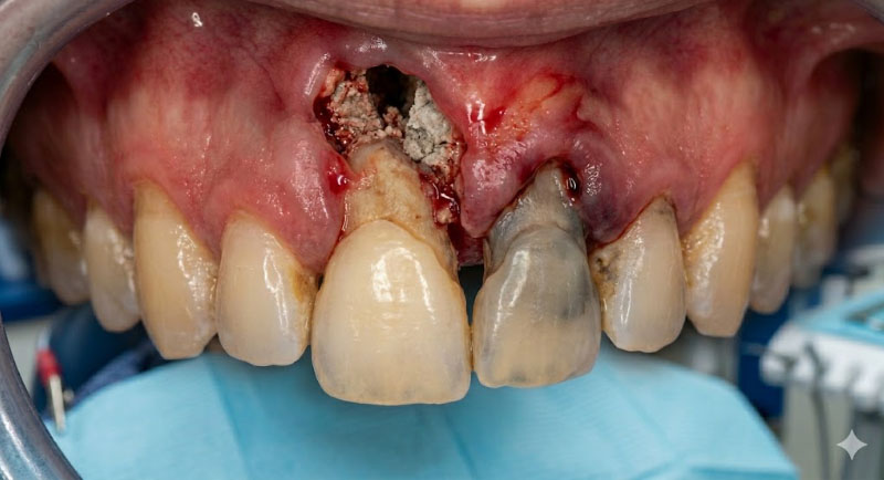
3. Exposure of the Graft Through the Gum
After bone grafting, the gum tissue should protect the surgical site while it heals. In some cases, the incision opens or the covering membrane becomes exposed.
A small amount of visible graft material does not automatically establish that the entire graft has failed. However, exposure can increase the risk of contamination and interfere with healing.
Warning signs
-
Gum tissue separating along the incision.
-
Visible graft particles or a membrane.
-
Persistent bleeding or discharge.
-
Increasing pain, swelling, or foul taste.
-
A wound that does not appear to be closing as expected.
Patients should not push graft particles back into the wound, pull on a membrane, or repeatedly touch the area with their fingers or tongue.
The treating clinician will determine whether observation, local wound care, additional protection, or surgical intervention is appropriate.
4. Sinus Complications After Upper-Jaw Bone Grafting
Bone grafting in the upper back jaw may be performed close to the maxillary sinus, an air-filled cavity above the upper teeth.
During certain procedures, including sinus augmentation, the sinus membrane can be torn or a communication can develop between the mouth and sinus. These complications vary in severity and do not always result in permanent damage.
Symptoms that need assessment
-
Persistent or worsening pressure or pain in the cheek.
-
Unusual nasal discharge or recurrent sinus symptoms after surgery.
-
Fluid passing between the mouth and nose.
-
Air passing through the surgical area.
-
Persistent symptoms that suggest an opening between the mouth and sinus.
Fluid passing between the mouth and nose after upper-jaw surgery warrants prompt contact with the treating surgeon. Do not test for an opening by forcefully blowing your nose.
If a sinus lift or other sinus-adjacent procedure has been performed, follow the surgeon's specific instructions. These may include avoiding nose blowing, sneezing with the mouth open, and avoiding pressure-generating activities for the prescribed period.
Persistent sinus complications may require medication, surgical closure, or revision of the graft, depending on the diagnosis.
5. Nerve Injury or Persistent Numbness
Some bone graft procedures take place near nerves that supply sensation to the lower lip, chin, teeth, tongue, or other oral structures.
Temporary altered sensation can occur after surgery, but persistent or new numbness requires assessment, particularly if it involves the lower lip, chin, or tongue.
Symptoms to report
-
Numbness that lasts longer than the surgeon advised.
-
New tingling, burning, or electric-shock-like sensations.
-
Persistent altered sensation after the local anesthetic should have worn off.
-
Changes in taste or sensation affecting the tongue.
The extent of nerve involvement cannot be determined from symptoms alone. The clinician may assess sensation and review imaging to determine whether observation, further investigation, or specialist management is needed.
Permanent sensory changes are uncommon but possible. Patients should report concerning symptoms early rather than waiting several weeks without advice.
6. Severe Bleeding or an Expanding Hematoma
Some blood staining and mild oozing may occur after oral surgery. Persistent heavy bleeding, however, requires immediate attention.
What should patients do?
If bleeding occurs, sit upright and apply firm, continuous pressure with clean gauze directly over the surgical area, if this is feasible and consistent with the surgeon's instructions. Avoid repeatedly lifting the gauze to check the wound.
If bleeding remains heavy or does not stop with sustained pressure, seek urgent dental or medical care. Call emergency services for uncontrolled bleeding, fainting, breathing difficulty, or signs of significant blood loss.
Patients taking anticoagulants or antiplatelet medicines should tell their surgeon about all medications before surgery. They should not stop prescribed blood-thinning medication without guidance from the prescribing clinician.
7. Delayed Healing and the Need for Repeat Surgery
Bone graft healing takes time. A graft may need several months to mature before an implant can be placed, and the exact interval depends on the grafting technique, the amount of bone being rebuilt, and the patient's healing response.
Delayed healing does not necessarily mean permanent failure. However, persistent inflammation, wound breakdown, infection, or insufficient bone volume can change the expected treatment schedule.
In some cases, a second grafting procedure is necessary. The clinician should explain why the original result is inadequate, what can be changed in the revised plan, and whether alternative options are available.
Patients should be cautious about promises of guaranteed graft success or a fixed implant-placement date before the site has been clinically reassessed.
8. Who May Have a Higher Risk of Complications?
Risk varies from patient to patient. Factors that deserve discussion before surgery include:
-
Smoking and nicotine exposure: These can impair healing and increase the risk of graft complications.
-
Poorly controlled diabetes: This can affect wound healing and infection risk.
-
Active oral infection: Existing infection may need to be treated before reconstruction.
-
Complex or extensive bone defects: Larger reconstructions can require more involved surgery and recovery.
-
Certain medications or medical conditions: These may affect bleeding, immune function, or bone healing.
-
Poor postoperative wound protection: Early trauma, pressure, or failure to follow the surgeon's instructions may compromise healing.
These factors do not mean that a patient will necessarily experience graft failure. They help the clinician assess risk and individualize the treatment plan.
Patients should provide a complete medical and medication history, follow postoperative instructions, maintain the recommended oral hygiene routine, and attend scheduled reviews.
9. When Is It a Dental Emergency?
Knowing when to seek help is one of the most important parts of safe recovery.
Seek emergency medical care immediately if you experience:
-
Difficulty breathing or swallowing.
-
Rapidly spreading swelling of the face, floor of the mouth, or neck.
-
Severe swelling associated with systemic illness or rapidly worsening symptoms.
-
Uncontrolled bleeding.
-
Collapse, confusion, or other signs of serious illness.
Contact your oral surgeon urgently for increasing pain, pus, fever, wound opening, persistent numbness, or suspected communication between the mouth and sinus.
Do not wait for a routine appointment if symptoms are rapidly worsening.
10. Expert Case-Analysis Approach: How Dentists Assess a Suspected Graft Complication
When a patient reports that a bone graft feels wrong, a careful clinical assessment should establish the likely cause before treatment is selected.
Step 1: Review the symptom timeline
The clinician asks when the surgery occurred, whether symptoms initially improved, and whether pain, swelling, bleeding, or discharge has changed.
Step 2: Examine the surgical site
The gum closure, wound integrity, tenderness, discharge, graft exposure, and surrounding tissues are assessed. The patient's temperature and general condition may also be relevant.
Step 3: Review imaging when indicated
Existing radiographs or additional imaging, including cone-beam computed tomography (CBCT) when justified, may help assess the bone defect, graft area, implant position, or nearby anatomical structures. Imaging findings must be interpreted alongside the clinical examination.
Step 4: Determine whether the graft is salvageable
The clinician evaluates whether the problem can be managed conservatively or whether drainage, debridement, graft removal, or additional reconstruction is required.
Step 5: Revise the treatment plan
The patient should understand the diagnosis, available options, expected healing period, likely additional costs, and risks of proceeding with or delaying implant placement.
Final expert perspective
The worst-case scenario after dental bone grafting is not simply that the graft fails. It is that a serious complication—such as a spreading infection, uncontrolled bleeding, or a significant sinus or nerve problem—is overlooked and treatment is delayed.
The safest approach is timely clinical assessment, appropriate imaging when needed, individualized treatment, and clear follow-up instructions. A successful outcome depends on more than the graft material itself: surgical planning, the patient's health, wound protection, and monitoring all matter.
Medical information note: This article provides general educational guidance and is not a substitute for an examination by a licensed dentist or oral and maxillofacial surgeon.
Worst-Case Scenario #1: Graft Failure
The most common major complication is graft failure.
What Is Graft Failure?
Graft failure occurs when the bone graft does not successfully integrate with your jawbone.
Instead of developing into healthy living bone, the graft may:
- Break down prematurely
- Become infected
- Fail to receive adequate blood supply
- Be rejected by surrounding tissues
When this happens, the intended bone growth never occurs.
Why Is Graft Failure a Problem?
Without successful integration:
- Bone volume remains inadequate
- Dental implants cannot be placed safely
- Additional surgeries may become necessary
- Treatment timelines become significantly longer
Patients often become frustrated because they expected the graft to solve the problem permanently.
Instead, another surgical procedure may be required after healing.
Symptoms of Graft Failure
Potential warning signs include:
- Persistent pain
- Delayed healing
- Swelling that does not improve
- Gum tissue breakdown
- Exposure of graft particles
- Continued bone loss
Fortunately, many graft failures can be corrected with additional treatment.
Worst-Case Scenario #2: Severe Bone Infection (Osteomyelitis)
One of the most serious complications involves infection spreading into the jawbone itself.
This condition is known as osteomyelitis.
What Is Osteomyelitis?
Osteomyelitis occurs when bacteria invade bone tissue and establish a deep infection.
Unlike a minor gum infection, osteomyelitis can be difficult to eliminate because bacteria become protected within the bone structure.
Why Is It Serious?
A bone infection can:
- Destroy healthy bone tissue
- Delay healing dramatically
- Threaten neighboring teeth
- Cause significant pain
- Spread to nearby anatomical structures
In severe cases, portions of infected bone may need surgical removal.
Treatment for Osteomyelitis
Management often includes:
- Surgical cleaning
- Removal of infected tissue
- Long-term antibiotics
- Careful monitoring
Although this complication is uncommon, it represents one of the most serious potential outcomes of dental surgery.
Worst-Case Scenario #3: Nerve Damage
Nerve injury is one of the complications patients fear most.
Fortunately, permanent nerve damage is rare.
Why Does It Happen?
Certain nerves run through the jawbone.
In the lower jaw, the mandibular nerve provides sensation to:
- The lower lip
- Chin
- Lower teeth
- Portions of the tongue
If surgery occurs too close to these structures, irritation or injury may occur.
Symptoms of Nerve Injury
Possible symptoms include:
- Numbness
- Tingling
- Burning sensations
- Altered feeling
- Partial loss of sensation
Most cases improve over time.
However, severe injuries may result in long-term or permanent changes in sensation.
Modern Prevention Methods
Today's dentists use advanced technology to reduce risk.
These tools include:
- Digital X-rays
- Cone beam CT scans
- Surgical guides
- Three-dimensional planning
Careful planning dramatically reduces the likelihood of nerve complications.
Worst-Case Scenario #4: Sinus Complications
Upper jaw bone grafts require special consideration because of their proximity to the maxillary sinuses.
What Happens During a Sinus Lift?
When bone is insufficient in the upper back jaw, a sinus lift may be performed.
The surgeon carefully elevates the sinus membrane and places graft material underneath it.
This creates additional bone volume for future implants.
Potential Problems
Complications may include:
- Sinus membrane perforation
- Infection
- Chronic sinusitis
- Graft displacement
If graft material migrates into the sinus cavity, additional treatment may be necessary.
Signs of Sinus Complications
Patients may notice:
- Nasal congestion
- Facial pressure
- Persistent drainage
- Sinus headaches
- Difficulty breathing through the nose
Prompt treatment generally prevents long-term problems.
Worst-Case Scenario #5: Wound Breakdown (Dehiscence)
One of the most frustrating complications is dehiscence.
What Is Dehiscence?
Dehiscence occurs when the gum tissue covering the graft opens prematurely.
Instead of remaining sealed during healing, the tissue separates and exposes the graft underneath.
Why Is Exposure Dangerous?
Once exposed:
- Bacteria can enter the site
- Infection risk increases
- Healing slows
- Graft stability decreases
Large exposures may lead to partial or complete graft loss.
Causes of Dehiscence
Common causes include:
- Smoking
- Trauma from chewing
- Poor oral hygiene
- Excessive pressure on the site
- Thin gum tissue
- Aggressive brushing
Careful post-operative care greatly reduces this risk.
Related Clinical Cases
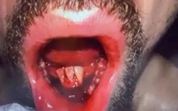
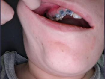
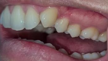
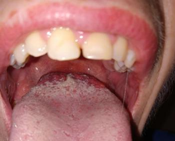
Patients with lower front gum inflammation may also experience tartar buildup, periodontal disease, gum abscesses, or tooth decay. Explore these related clinical case studies.