


Why Bone Grafts Fail in Some Dental Clinics: Risks, Causes, and Patient Advice

Topics:
Bone grafting is a common procedure used in implant dentistry to rebuild jawbone that has been lost due to tooth extraction, gum disease, or trauma. A successful bone graft creates enough bone volume to support a dental implant securely.
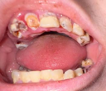
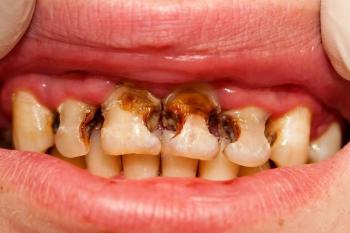
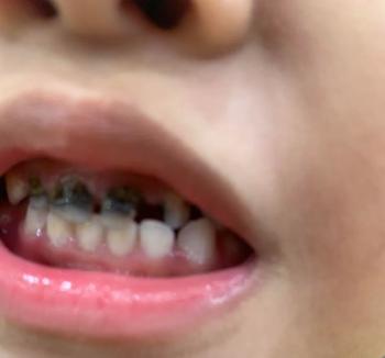
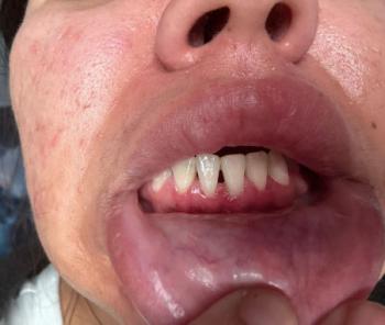
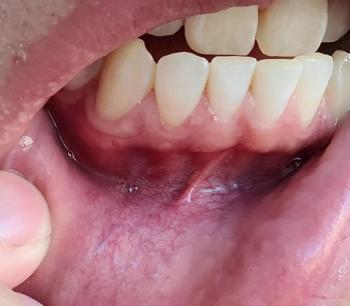
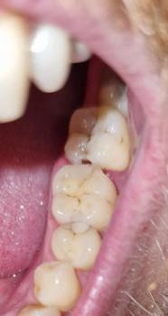
However, in some cases bone grafts fail. Patients may experience infection, poor bone formation, or graft rejection. One concern sometimes discussed in dental communities is that some clinics may perform bone graft procedures even when the patient is not an ideal candidate, often because the procedure is profitable.