
Dental Implants Cost Australia vs Philippines: The Hidden Truth Behind Saving Money Overseas
Topics:
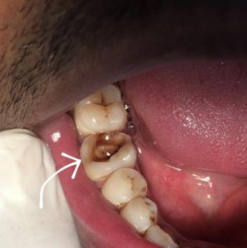
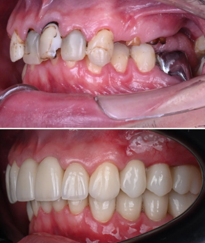
I Thought One Missing Tooth Didn't Matter — Until It Changed My Smile, My Confidence, and My Wallet
Have you ever lost a tooth and told yourself:
"It's only one tooth."
"Nobody can see it anyway."
"I'll replace it later when I have more money."
"I'm too busy right now."
If so, you're not alone.