


Root Canal Treatment in Connecticut, USA: How a Skilled Dentist Rescued a Tooth That Seemed Impossible to Save

When Everything Looked Lost
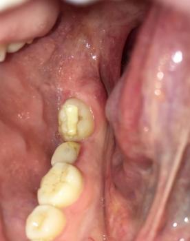
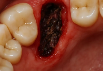
It was a cold winter morning in Connecticut when Michael, a 47-year-old construction supervisor, walked into a dental office with a look of exhaustion and pain. For nearly two weeks, he had been unable to sleep properly. The throbbing pain coming from his lower molar had become unbearable.
"I think the tooth is gone," he told the dentist.