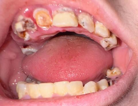
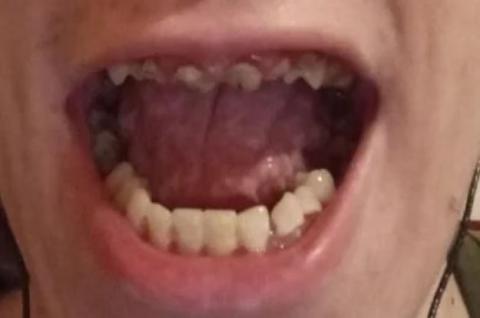
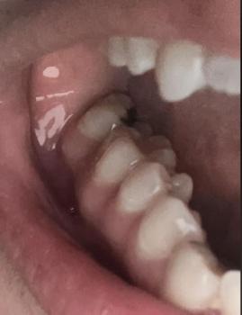
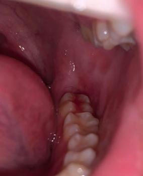
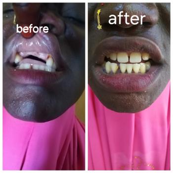
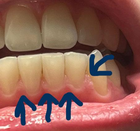
Gum Recession on Lower Front Teeth | Causes, Treatment, and Prevention
Severity:
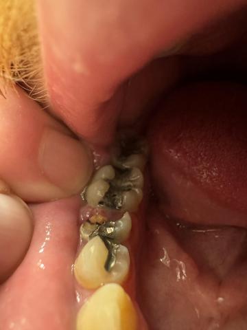
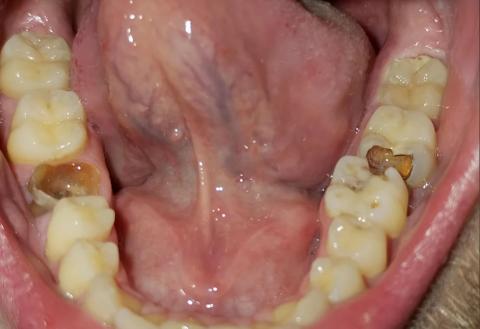
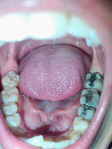
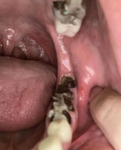
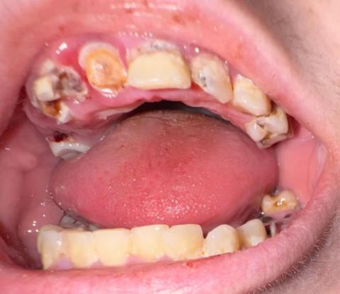
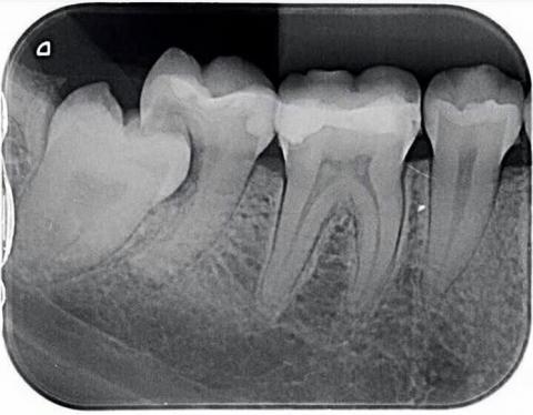
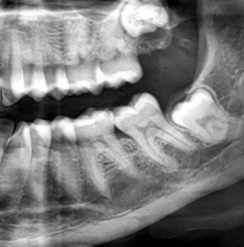
Teeth Problems:
Gum Recession and Cervical Tooth Wear on Lower Front Teeth – Full Dental Case Analysis (100% Zoom)
Medical Disclaimer
This analysis is image-based and for educational purposes only. A definitive diagnosis requires an in-person dental examination, periodontal probing, and dental X-rays. The findings below are based on visible clinical signs and standard dental guidelines.