Tooth decay

Lower Teeth with Amalgam Fillings Case Analysis

Severity: 

Dental Case Analysis: Lower Teeth with Metallic Fillings and Plaque Accumulation

Case Overview

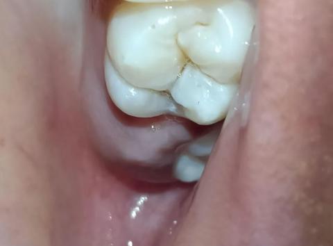
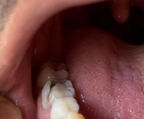
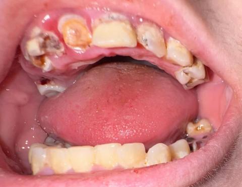
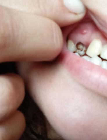
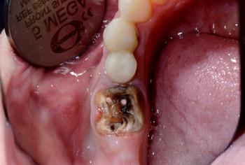
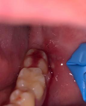
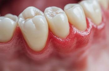
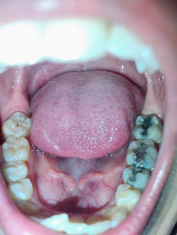
The image shows the lower dental arch with visible metallic (amalgam) restorations on posterior molars. There are signs of plaque accumulation and dark staining within the grooves of treated teeth.


Zoom 100% Visual Examination

Observed Findings

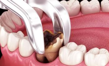
  • Multiple amalgam (silver) fillings on lower molars

  • Dark discoloration around restoration margins

Back Molar with Deep Fissures Case Analysis

Severity: 

Dental Case Analysis: Back Molar with Deep Grooves and Early Decay Risk

Case Overview

The image shows a posterior molar tooth with deep occlusal grooves (fissures). While the enamel surface appears mostly intact, the depth of the grooves creates a high risk area for early tooth decay due to plaque and food retention.


Zoom 100% Visual Examination

Observed Findings

  • Back molar with pronounced pits and fissures

Broken Back Tooth with Deep Cavity Case Analysis

Severity: 

Dental Case Analysis: Broken Back Tooth with Deep Cavity

Case Overview

The image shows a posterior tooth located near the tongue side with visible enamel breakdown and a deep cavity. The tooth structure appears weakened, suggesting advanced decay and possible fracture.


Zoom 100% Visual Examination

Observed Findings

  • Back tooth with broken enamel edge

  • Deep cavity extending into dentin

Severe Tooth Decay and Gum Infection Case Analysis

Severity: 

Dental Case Analysis: Multiple Advanced Tooth Decay With Gum Inflammation

Case Overview (100% Zoom Visual Inspection)

The image shows the upper and lower dental arches with multiple teeth affected by advanced decay, visible dark cavities, broken tooth structure, and inflamed gum tissue. Several teeth show severe enamel and dentin destruction, indicating a high-risk oral condition requiring urgent dental care.

Severe Tooth Decay With Gum Infection - Dental Case Analysis

Severity: 

Dental Case Analysis – Severe Tooth Decay With Localized Gum Infection

Case Overview

This intra-oral image shows a tooth with advanced decay and an associated localized gum swelling above the affected tooth. The appearance suggests an active dental infection likely originating from deep caries that has reached the pulp and spread toward the surrounding soft tissue.


Full Analysis and Diagnosis

Clinical Findings

  • Deep cavity with visible tooth structure breakdown

Pages