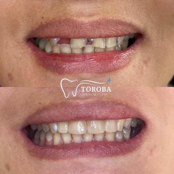
Your Smile, Our Passion.
Interproximal Tooth Decay Seen on Dental X-ray - Zoomed Case Analysis
Image:
Severity:
Teeth Problems:
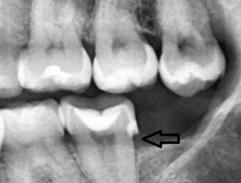
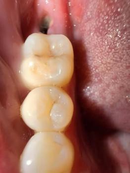
Teeth Case – Zoom 100% (Dental X-ray)
Focused Area: Posterior teeth (molar region) with interproximal defect indicated by arrow
Full Analysis and Deep Radiographic Examination
This assessment is based on the provided dental X-ray image only and does not replace a complete clinical examination, periodontal probing, or additional radiographs if required.
Radiographic Findings
-
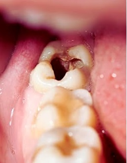
Radiolucent area between posterior teeth, consistent with interproximal dental caries
-
Possible loss of tooth structure at the contact point
-
Early signs of localized bone level reduction adjacent to the affected tooth
-
Existing restoration visible on the molar, suggesting previous decay history
-
No obvious advanced periapical abscess visible at this image depth, but early pathology cannot be ruled out
Probable Diagnosis
-
Interproximal dental caries (moderate depth)
-
Early periodontal bone involvement localized to the molar region
-
Increased risk of pulp irritation depending on cavity depth
Recommended Treatment Process
Step 1: Full Mouth Scaling and Polishing (Day 1)
-
Ultrasonic scaling to remove plaque and calculus
-
Manual scaling in posterior and interproximal areas
-
Polishing to reduce bacterial reattachment
Estimated duration: 45–60 minutes
Local anesthesia: May be required depending on sensitivity
Step 2: Definitive Interproximal Treatment (Day 1–7)
Based on X-ray depth and clinical findings:
-
Composite restoration for moderate interproximal decay
-
Replacement of failing restoration if margins are compromised
-
Root canal treatment if pulp involvement is confirmed
-
Crown placement if structural integrity is reduced
Healing and Recovery Timeline
| Time Frame | Expected Outcome |
|---|---|
| Days 1–3 | Gum irritation decreases after scaling |
| Days 4–7 | Stabilization of gingival tissues |
| Days 8–14 | Healthy gum response if decay is treated |
A 14-day healing period is realistic for soft tissue once infection sources are removed.
What Will Scale Up If Left Untreated
-
Expansion of interproximal decay into the pulp
-
Development of dental abscess or chronic infection
-
Progressive bone loss around the affected molar
-
Increased tooth mobility and eventual tooth loss
-
Higher cost and complexity of future dental treatment
Post-Treatment Care Instructions
-
Brush twice daily using fluoride toothpaste
-
Floss daily, especially between molars
-
Use interdental brushes if contacts are open
-
Maintain regular dental checkups and X-rays as advised
Professional Comment
Radiographic interproximal decay often progresses silently without pain. Scaling alone will not stop this condition. Early restorative treatment guided by X-ray findings is critical to preserve the tooth and surrounding bone.
Visit a Dental Clinic Near You
For dental X-rays, scaling, and interproximal cavity treatment, locate a nearby dental clinic using our directory:
Teeth Case
 |
 |
 |
 |
 |
 |