Xray

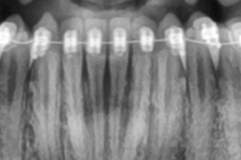
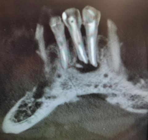
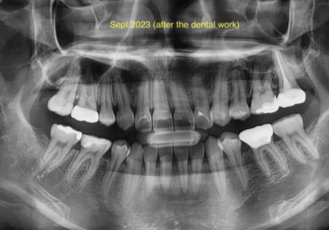
Panoramic X-Ray Showing Generalized Bone Loss Case Analysis

Severity: 
Teeth Problems: 

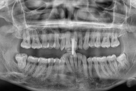
Panoramic X-Ray Showing Generalized Bone Loss and Infection Risk Case Analysis

What Is Seen in This Case

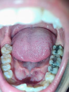
The panoramic X-ray shows both upper and lower jaws with multiple teeth present, but the bone levels around many tooth roots appear reduced. In several areas, the bone looks uneven and lower than normal, especially around the front teeth and molar regions. There is also evidence of previous dental treatment on at least one tooth.

Bone Loss Around Front Teeth With Braces X-Ray Case

Severity: 
Teeth Problems: 

Bone Loss Around Front Teeth With Braces X-Ray Case Analysis

What Is Seen in This Case

The X-ray image shows the lower front teeth with orthodontic braces in place. The tooth roots are visible, but the bone level around the roots appears reduced and uneven. There are darker areas between and around the roots, suggesting loss of supporting bone rather than healthy, dense bone.

This finding is especially important in patients undergoing orthodontic treatment.

Severe Bone Infection Around Tooth Roots X-Ray Case

Severity: 

Severe Bone Infection Around Tooth Roots X-Ray Case Analysis

What Is Seen in This Case

The X-ray image shows severe bone loss and dark radiolucent areas around the roots of multiple teeth. The surrounding jawbone appears irregular, hollowed, and damaged, which strongly indicates advanced infection spreading through the bone.

The tooth roots are clearly visible with loss of normal bone support, a serious sign that the infection is no longer limited to the gums.

Dental X-Ray Showing Bone Infection and Abscess Case Analysis

Severity: 

What Is Seen in This Case

The dental X-ray shows bone changes around the roots of the teeth, particularly in the back tooth area. There are visible signs of bone loss and darkened areas near the root tips, which commonly indicate a tooth abscess or chronic dental infection.

Some teeth appear previously treated, but the surrounding bone suggests ongoing or unresolved infection.


Full Analysis and Provisional Diagnosis

Based on the X-ray appearance, the most likely findings include:

Panoramic Dental X-Ray Case Analysis After Dental Treatment

Severity: 

What Is Seen in This X-Ray

This panoramic dental X-ray (taken after dental work in September 2023) shows multiple restored teeth, including fillings and treated posterior teeth. The roots and surrounding bone are visible across the upper and lower jaws, allowing evaluation of healing, hidden infection, and overall oral health.

Some areas show treated teeth with restorations, while other regions require close monitoring for possible residual or recurrent infection.

Pages