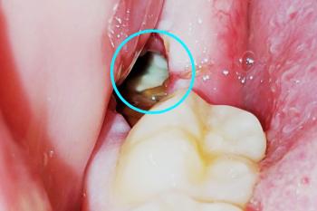
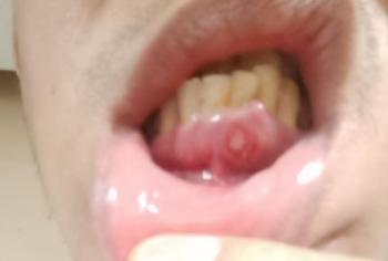
Molar Tooth Early Decay and Gum Pocket Case Analysis
Severity:
Teeth Problems:
Dental Case Analysis: Molar Tooth with Early Decay and Gum Pocket
Case Overview
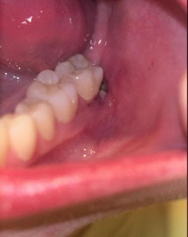
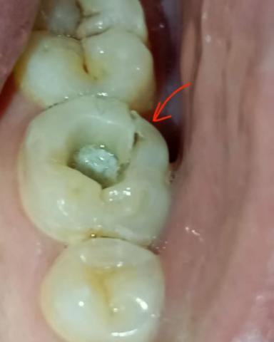
The image shows a back molar tooth with a small occlusal cavity and a visible gum-side pocket or irritation (indicated by the arrow). This suggests early tooth decay combined with localized gum involvement.
Zoom 100% Visual Examination
Observed Findings
-
Posterior molar with small cavity on the chewing surface
-
Early enamel breakdown visible in the fissure