Where Bright Smiles Begin.

Root Canal Infection With Apical Bone Loss - Dental Case Analysis
Image:
Severity:
Teeth Problems:
Understanding Root Canal Infection and Apical Bone Loss: Expert Dental Guidance
A tooth that develops an infection around its root after root canal treatment may still be treatable. However, persistent infection and loss of supporting bone require a careful dental examination to determine whether the tooth can be saved and which treatment is most appropriate.
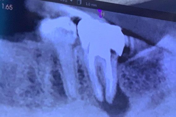
In this dental case, the X-ray description identifies a previously root canal-treated posterior tooth with a dark area around the root tip, reduced normal bone pattern, and possible widening of the periodontal ligament space. These findings can be consistent with apical periodontitis, an inflammatory condition affecting the tissues surrounding the root tip.
A dark area on an X-ray does not, by itself, prove that bacteria are actively spreading or establish the exact cause. The dentist must interpret the image alongside the patient's symptoms, dental history, examination findings, and other relevant tests.
1. What Causes Infection After Root Canal Treatment?
Root canal treatment removes infected or inflamed pulp tissue, cleans and disinfects the root canal system, and seals the canals to help prevent further bacterial contamination. Although the procedure is generally successful, some teeth may develop persistent or recurrent disease.
Possible causes include:
-
Missed root canals: Complex root anatomy may leave untreated spaces where bacteria can persist.
-
Incomplete cleaning or sealing: Difficult canal anatomy or technical limitations may prevent adequate treatment.
-
Leakage around a filling or crown: A defective restoration or new tooth decay may allow bacteria to re-enter the tooth.
-
Cracks or fractures: A damaged tooth can provide a pathway for bacterial contamination and may affect whether the tooth can be saved.
-
Persistent inflammation: Bone and surrounding tissues may take time to heal, and some lesions do not resolve as expected.
A previously treated tooth can therefore develop problems months or even years after the original procedure. An X-ray abnormality should be assessed rather than automatically classified as a failed root canal.
2. What Does Apical Bone Loss Mean?
Apical bone loss refers to the loss of bone around the tip of a tooth's root. When bacteria from an infected or necrotic root canal stimulate inflammation in the surrounding tissues, the body's immune response can cause localized changes in the supporting bone.
On a dental X-ray, this process may appear as a radiolucency, or a darker area, near the root apex.
Depending on the clinical findings, possible explanations include:
-
Chronic apical periodontitis.
-
A persistent or recurrent periapical inflammatory lesion.
-
A developing or established apical abscess, if supported by clinical signs.
-
Other conditions that can resemble an inflammatory lesion on imaging.
Important clinical distinction: Not every dark area at the root tip is an abscess, and not every radiographic lesion requires immediate extraction. The diagnosis determines the appropriate treatment.
3. Can a Tooth With Apical Bone Loss Still Be Saved?
Yes, in many cases. The presence of bone loss does not automatically mean that the tooth must be removed.
An endodontist or appropriately trained dentist will assess the remaining tooth structure, periodontal support, root anatomy, previous treatment, and the extent of the lesion.
Potential treatment options include:
Nonsurgical root canal retreatment
The dentist reopens the tooth, removes the existing root canal filling material when appropriate, identifies missed anatomy, disinfects the canals, and reseals them. This is often considered when the original root canal treatment can be corrected and the tooth is restorable.
Endodontic microsurgery
An apicoectomy may be considered when infection persists despite appropriate root canal treatment or when conventional retreatment is unsuitable. The procedure involves accessing the root tip, removing affected tissue as indicated, and sealing the root end.
Extraction
Removal may be necessary if the tooth has a non-restorable fracture, insufficient remaining structure, severe periodontal damage, or an unfavorable prognosis after considering available treatment options.
When the tooth is suitable for preservation, the dentist should discuss the benefits, limitations, cost, and expected prognosis of retreatment before recommending extraction.
4. How Long Does Apical Bone Healing Take?
Healing varies with the original diagnosis, lesion size, treatment quality, the patient's general health, and other clinical factors.
Some patients notice improvement in pain or tenderness within days after appropriate treatment. However, symptom improvement does not necessarily mean that the bone has completely healed.
Radiographic healing commonly takes several months and can continue for a year or longer. Some lesions take longer to resolve, and not all lesions heal completely.
Follow-up appointments and comparison with previous X-rays help the dentist determine whether the lesion is shrinking, remaining stable, or progressing. A fixed healing deadline should not be promised for every patient.
5. Are Antibiotics Enough to Cure a Root Canal Infection?
Usually, no. Antibiotics cannot reliably eliminate the source of infection inside a necrotic root canal. Definitive dental treatment is needed to address the cause.
Depending on the diagnosis, this may involve root canal treatment or retreatment, drainage of an abscess, or extraction of a tooth that cannot be saved.
Antibiotics may be indicated when there is systemic involvement, such as fever or malaise, or when specific clinical circumstances warrant their use. They should be prescribed by a qualified clinician following an individual assessment.
Do not take leftover antibiotics, share medication, or delay necessary dental treatment while waiting for antibiotics to work.
6. Warning Signs That Require Urgent Attention
Arrange a prompt dental examination if you experience persistent tooth pain, pain when biting, gum swelling near a treated tooth, drainage, a recurring gum pimple, or a bad taste associated with the area.
Seek emergency medical care immediately if you develop difficulty breathing or swallowing, rapidly spreading facial or neck swelling, or swelling that affects the eye or vision. Fever with increasing facial swelling or feeling seriously unwell also warrants urgent assessment.
An infection can occasionally spread beyond the tooth and surrounding bone. Do not wait for a routine follow-up appointment if these warning signs appear.
Expert Clinical Takeaway
Apical bone loss around a previously root canal-treated tooth is an important finding, but it is not sufficient on its own to determine the tooth's prognosis. A dentist must identify the likely source of the lesion, evaluate the restorability of the tooth, and select treatment based on the complete clinical picture.
Early assessment can help prevent further complications and may preserve a natural tooth when appropriate treatment is feasible.
Frequently Asked Questions About Root Canal Infection and Bone Loss
1. Can a root canal-treated tooth become infected again?
Yes. Persistent or recurrent infection can occur if bacteria remain in untreated canal anatomy, if the tooth becomes contaminated through a leaking restoration, or if a new problem such as decay or fracture develops. A dentist can assess whether retreatment or another intervention is appropriate.
2. Does bone loss around a root canal-treated tooth mean the treatment failed?
Not necessarily. Some lesions may represent persistent disease, while others may be healing slowly after treatment. Comparing previous and current X-rays, reviewing symptoms, and performing a clinical examination help distinguish these possibilities.
3. Can apical bone loss grow back after treatment?
Yes. When the source of inflammation is adequately controlled, the surrounding bone can gradually repair and remodel. The amount and speed of healing vary, and follow-up imaging is needed to evaluate progress.
4. Can a tooth have an infection without pain?
Yes. Some chronic apical lesions cause few or no symptoms and may be discovered during routine dental imaging. The absence of pain does not rule out disease, so unexplained changes around a root tip should be evaluated.
5. Is root canal retreatment better than extraction?
Neither option is best for every patient. Retreatment may be appropriate when the tooth is structurally sound enough to restore and has a reasonable chance of healing. Extraction may be more appropriate for a severely fractured or non-restorable tooth. The decision should follow an individualized prognosis assessment.
6. What is the difference between apical periodontitis and a dental abscess?
Apical periodontitis is inflammation of the tissues around the root tip, often associated with infection inside the root canal. A dental abscess is a localized collection of pus. An abscess may develop in association with apical infection, but a radiographic lesion alone does not establish that pus is present.
7. Will antibiotics remove the dark spot on my dental X-ray?
Antibiotics alone are not a dependable treatment for the underlying cause of most infections within a necrotic root canal. When the infection source is controlled, the bone may gradually heal, and the radiographic lesion may become smaller. Healing should be assessed over time.
8. How often should a dentist monitor apical bone loss?
The schedule depends on the diagnosis, symptoms, treatment performed, and initial lesion characteristics. A dentist may arrange an earlier clinical review and subsequent radiographs over several months or longer. Additional imaging should be guided by clinical need rather than a rigid schedule.
9. Can a large apical lesion still heal without extraction?
Potentially, yes. Lesion size alone does not determine whether a tooth can be saved. The cause of the lesion, tooth restorability, periodontal condition, root anatomy, and response to treatment all influence the prognosis.
10. When should I see an endodontist?
Consider referral when a previously treated tooth has a persistent or enlarging apical lesion, recurrent symptoms, complicated root anatomy, suspected missed canals, or a possible need for endodontic surgery. An endodontist can help evaluate tooth-preserving options and their likely outcomes.
Medical disclaimer: This educational case analysis is not a substitute for an in-person dental examination or a diagnosis from a qualified dentist. Treatment decisions should be based on the patient's clinical findings and appropriate imaging.
Dental Case Analysis – Root Canal Infection With Apical Bone Loss
Case Overview
This dental X-ray shows a posterior tooth with previous root canal treatment. A clear radiolucent area at the root apex is visible, indicating a persistent or recurrent infection. The surrounding bone shows signs of loss consistent with chronic inflammation.
Full Analysis and Diagnosis
Radiographic Findings
-
Root canal–treated tooth with dense coronal restoration
-
Apical radiolucency at the root tip
-
Loss of normal bone trabeculation
-
Widened periodontal ligament space
-
Infection appears chronic rather than acute
Tooth Condition
-
Root canal filling may be incomplete or leaking
-
Possible missed canal or bacterial persistence
-
Apical seal likely compromised
-
Tooth structure remains intact but weakened
Diagnosis
-
Chronic apical periodontitis
-
Failed or compromised root canal treatment
-
Localized periapical bone loss
Time Frame to Heal
With Proper Treatment
-
Infection control: 7–14 days
-
Reduction in inflammation: 1–2 weeks
-
Bone healing begins: 4–6 weeks
-
Radiographic bone regeneration: 3–6 months
Without Treatment
-
Infection persists or enlarges
-
Progressive bone destruction
-
Pain, swelling, or abscess formation
-
Increased risk of tooth loss
Process to Execute (14-Day Window)
Days 1–3
-
Clinical evaluation and X-ray review
-
Confirm infection source
-
Prescribe antibiotics if clinically indicated
Days 4–7
-
Root canal retreatment or
-
Surgical intervention (apicoectomy) if retreatment is not possible
-
Removal of infected tissue
Days 8–14
-
Post-treatment evaluation
-
Monitor symptoms and healing
-
Plan long-term restoration if needed
Issues That Will Scale Up If Untreated
-
Expansion of apical lesion
-
Further jawbone loss
-
Chronic pain or swelling
-
Sinus or nerve involvement (depending on location)
-
Eventual tooth extraction
Clinical Comments
This type of infection can remain silent for long periods and is often discovered only through imaging. Early intervention significantly improves the chance of saving the tooth and preventing further bone loss.
Find a Dental Clinic Near You
Search for a qualified dental clinic in your area:
https://cebudentalimplants.com/map-dental-clinic
Related Clinical Cases
Patients with lower front gum inflammation may also experience tartar buildup, periodontal disease, gum abscesses, or tooth decay. Explore these related clinical case studies.