Because Every Tooth Deserves Care.

Root Caries on Front Teeth | Cervical Tooth Decay Case Analysis and Treatment Guide
Image:
Severity:
Teeth Problems:
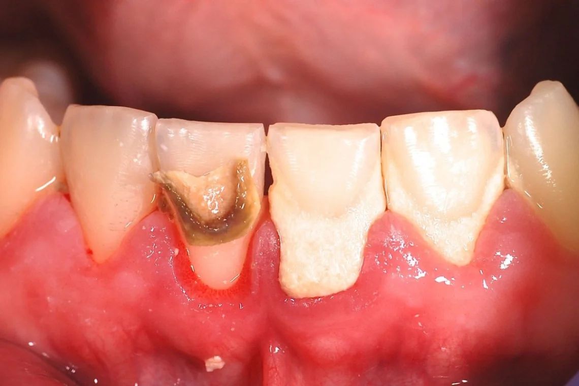
Advanced Cervical / Root Caries on Anterior Teeth – Full Case Analysis (100% Zoom)
Clinical Note: The following is an educational analysis based on the provided image. A definitive diagnosis requires an in-person dental examination, periodontal probing, and dental X-rays.
Case Overview (Zoom 100%)
The image shows localized but advanced decay at the cervical (gum-line) and root surface of an anterior tooth, with adjacent teeth also showing gingival recession. The darkened area indicates dentin/root involvement, not surface staining. The gum tissue appears inflamed and recessed, exposing softer root surfaces that are highly vulnerable to rapid decay.
Primary Diagnosis
-
Root Caries (Cervical Caries)
-
Decay has progressed into root cementum and dentin
-
Faster progression than enamel caries due to softer tooth structure
-
-
Gingival Recession
-
Gum tissue has receded, exposing the root
-
Commonly linked to periodontal disease, aggressive brushing, or occlusal stress
-
-
High Sensitivity / Early Pulp Risk
-
Lesion depth suggests proximity to the pulp
-
Risk of pulpitis if not treated promptly
-
Client-Reported Symptoms (Typical in This Case)
-
Sharp sensitivity to cold, air, or sweets
-
Occasional pain when brushing or eating
-
Gum tenderness or bleeding near the affected tooth
-
Concern about tooth appearance or dark discoloration
Deep Examination Findings (Image-Based)
-
Clearly demarcated carious lesion at the root surface
-
Loss of normal tooth contour near the gum line
-
Inflamed gingiva with reduced attachment height
-
Adjacent teeth show early risk due to recession
Can This Heal in 14 Days?
Tooth decay does NOT heal on its own.
However, 14 days is a critical window to:
-
Stop bacterial progression
-
Reduce inflammation
-
Prevent nerve infection
Without treatment, damage will continue beyond this period.
Treatment Process to Execute
Step 1: Clinical Assessment (Day 1–3)
-
Intraoral examination
-
Periapical X-ray of affected tooth
-
Periodontal evaluation
Step 2: Disease Control (Day 3–7)
-
Scaling and root planing to remove plaque and bacteria
-
Desensitizing agents if needed
-
Oral hygiene correction (brushing technique, fluoride use)
Step 3: Definitive Restoration (Day 7–14)
Depending on severity:
-
Cervical composite filling (if pulp not involved)
-
Root canal treatment (if pulp is infected)
-
Crown placement if structural integrity is compromised
-
Extraction only if the tooth is non-restorable
Expected Healing Timeline
-
Gum inflammation reduction: 7–10 days
-
Sensitivity improvement: 1–2 weeks
-
Full soft tissue stabilization: 3–4 weeks
-
Long-term stability depends on oral hygiene and recall visits
What Will Scale Up If Left Untreated
-
Rapid deepening of root decay
-
Severe tooth sensitivity and pain
-
Pulp infection and abscess formation
-
Gum and bone loss around the tooth
-
Tooth fracture or eventual tooth loss
-
Increased treatment cost and complexity
Professional Comments
-
Root caries is aggressive and time-sensitive
-
Early treatment can save the tooth and nerve
-
Delay significantly increases the risk of root canal or extraction
-
Preventive care and regular follow-ups are essential after treatment
Strong Recommendation
Please visit the nearest dental clinic as soon as possible for a full evaluation and appropriate treatment.
You may search for a clinic near your location using our dental directory:
https://cebudentalimplants.com/map-dental-clinic
Early intervention can prevent pain, infection, and tooth loss.
Related Clinical Cases
Patients with lower front gum inflammation may also experience tartar buildup, periodontal disease, gum abscesses, or tooth decay. Explore these related clinical case studies.



Other topics:
Other Dental Clinic