Dentures That Feel Natural, Look Beautiful.
Periapical Radiolucency Explained: Causes, Diagnosis, and Fastest Treatment for Apical Infection
Image:
Severity:
Teeth Problems:
Periapical Radiolucency Case: Apical Infection, Bone Loss, and 14-Day Healing Guide
FULL ANALYSIS (X-RAY INTERPRETATION)
1. Radiolucent Lesion at the Apex
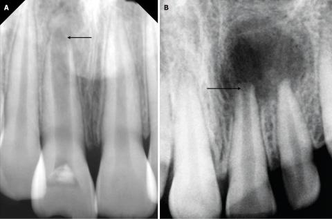
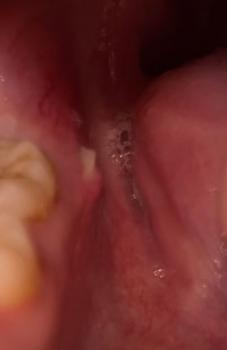
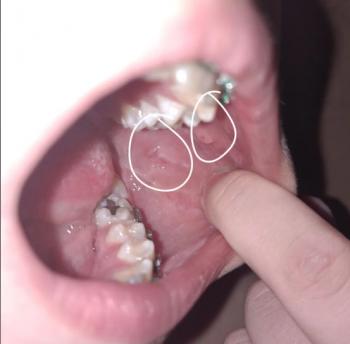
Both Image A and Image B show a dark, well-defined radiolucent area surrounding the root apex of one of the anterior teeth. This appearance is consistent with:
-
Periapical abscess
-
Periapical cyst
-
Chronic apical granuloma
-
Chronic apical periodontitis
2. Loss of Lamina Dura
The normal white cortical outline around the root tip is not clearly visible. Loss of the lamina dura typically indicates a non-vital pulp and chronic inflammation.
3. Widened Periodontal Ligament Space
This suggests ongoing inflammatory changes around the root.
4. Bone Rarefaction
The dark zone indicates bone loss caused by chronic infection. This usually results from long-standing pulp death or untreated decay or trauma.
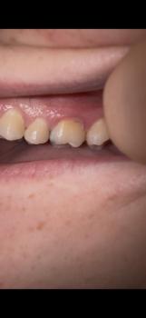
5. Tooth Structure
The visible crowns appear intact, but the root area strongly suggests the tooth is no longer vital.
TIME FRAME TO HEAL
Day 0 to 14
-
Symptoms reduce after treatment
-
Inflammation begins to subside
-
The socket or periapical area stabilizes
1 to 3 Months
-
Bone begins remodeling and filling in
-
The radiolucent area gradually decreases
6 to 12 Months
-
Noticeable bone regeneration
-
Radiograph shows significant shrinkage of the lesion
12 to 24 Months
-
Complete bone regeneration is possible
-
Radiolucency may disappear entirely on X-ray
IF AFTER 14 DAYS THERE IS NO IMPROVEMENT
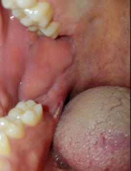
These complications may develop or worsen:
-
Infection may spread to surrounding tissues
-
Enlargement of a periapical cyst
-
Increased bone loss around the roots
-
Tooth may become non-restorable if left untreated
-
Infection may spread to adjacent teeth
-
Possible abscess formation or swelling
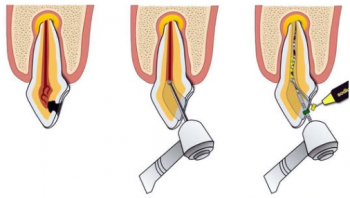
PROCESS TO EXECUTE
-
Conduct a pulp vitality test
-
Confirm diagnosis through clinical and radiographic examination
-
Perform root canal treatment if the tooth is non-vital
-
Prescribe antibiotics only if swelling, fever, or spreading infection is present
-
Schedule follow-up radiographs at 3, 6, and 12 months
-
If root canal treatment fails, consider apicoectomy or extraction
COMMENTS
The X-ray indicates a chronic apical lesion consistent with long-standing infection. Immediate dental management is recommended to prevent further bone loss. Early intervention has a high success rate, especially with properly performed root canal therapy.
NEAREST DENTAL LOCATION
You can locate the nearest dental clinic through:
https://cebudentalimplants.com/map-dental-clinic
Teeth Case
 |
 |
 |
 |
 |
 |