From Pain to Perfect — We Care for Your Smile.

Localized Gum Recession on Premolar | Causes, Treatment, and Prevention
Image:
Severity:
Teeth Problems:
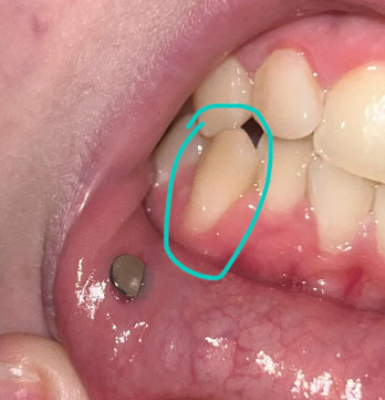
ocalized Gum Recession and Root Exposure on Lower Premolar – Full Dental Case Analysis (100% Zoom)
Medical Disclaimer
This analysis is image-based and for educational purposes only. A definitive diagnosis requires an in-person dental examination, periodontal probing, and dental X-rays. The findings below are based on visible clinical signs and accepted dental standards.
Case Overview (Zoom 100%)
The circled tooth shows localized gum recession on a lower premolar, with visible root surface exposure. The gum margin has migrated downward, exposing the lighter-colored root area compared to the enamel above. The surrounding gum tissue appears mildly inflamed but without obvious pus or acute infection.
A metal stud visible on the lip is unrelated to the dental condition.
This presentation is consistent with early-to-moderate gingival recession, not active tooth decay.
Primary Diagnosis
-
Localized gingival recession on a lower premolar
-
Exposed root surface (cementum/dentin exposure)
-
Early non-carious cervical lesion risk
-
Increased susceptibility to tooth sensitivity and root caries
Deep Clinical Examination (Image-Based Findings)
-
Gum margin positioned apical to its normal level
-
Smooth, light-colored exposed root surface
-
Mild gingival inflammation at the recession margin
-
No visible abscess, ulceration, or necrotic tissue
-
Likely contributing factors:
-
Aggressive or improper brushing technique
-
Thin gum biotype
-
Local plaque accumulation
-
Occlusal stress or tooth position
-
Can This Heal in 14 Days?
Gum recession does not reverse naturally.
However, within 14 days, appropriate dental care can:
-
Stop further recession
-
Reduce inflammation
-
Decrease or prevent sensitivity
-
Protect the exposed root surface
Early management is key to long-term stability.
Recommended Treatment Process
Step 1: Periodontal Assessment (Days 1–3)
-
Periodontal probing to measure recession depth
-
Evaluation of gum thickness and attachment level
-
Assessment of brushing habits and bite forces
Step 2: Disease Control and Prevention (Days 3–7)
-
Professional scaling and polishing
-
Removal of plaque near the gum line
-
Instruction on gentle brushing technique
-
Use of desensitizing toothpaste if sensitivity exists
Step 3: Protective or Corrective Care (Days 7–14)
Depending on severity and symptoms:
-
Fluoride or desensitizing varnish
-
Cervical composite restoration if root wear progresses
-
Occlusal adjustment if traumatic bite is present
-
Periodontal referral if recession continues to worsen
Expected Healing Timeline
-
Reduction in gum inflammation: 7–10 days
-
Sensitivity improvement: 1–2 weeks
-
Long-term stabilization depends on habit correction and maintenance
Note: Gum tissue will stabilize but will not regrow without surgical intervention.
What Will Escalate If Untreated
-
Increased tooth sensitivity
-
Higher risk of root caries
-
Progressive gum recession
-
Aesthetic concerns due to elongated tooth appearance
-
Potential future tooth mobility in advanced cases
Professional Comments
This is a common and manageable dental condition. When addressed early, most patients maintain their teeth long-term without surgery. The key is early prevention and habit correction, not aggressive treatment.
This is not an emergency, but it should not be ignored.
Strong Recommendation
Schedule a dental visit for periodontal evaluation and preventive treatment.
You can find a dental clinic near your area using this directory:
https://cebudentalimplants.com/map-dental-clinic
Early care prevents more complex and costly procedures later.
Related Clinical Cases
Patients with lower front gum inflammation may also experience tartar buildup, periodontal disease, gum abscesses, or tooth decay. Explore these related clinical case studies.



Other topics:
Other Dental Clinic