Dentures That Feel Natural, Look Beautiful.

Localized Gum Recession Around a Premolar: Expert Guidance for Exposed Tooth Roots
Image:
Severity:
Teeth Problems:
Understanding Why a Premolar Root Becomes Exposed
Localized gingival recession occurs when the gum margin moves away from its normal position, exposing part of the tooth root. When this affects a premolar, the tooth may appear longer than its neighbors, and the exposed root may become sensitive to cold drinks, brushing, or sweet foods.
Unlike the crown, the root surface is covered by cementum rather than enamel. This makes an exposed root more vulnerable to root-surface decay and wear. However, not every case of gum recession is caused by gum disease, and not every exposed root requires surgery.
A careful dental examination is essential to identify the cause, assess the amount of tissue loss, and determine whether the recession is stable or progressing.
What Causes Localized Premolar Gum Recession?
Several factors can contribute to recession around a single premolar:
-
Aggressive toothbrushing: Excessive pressure or a hard-bristled toothbrush may contribute to gum injury and tissue loss.
-
Periodontal disease: Inflammation and destruction of the tissues supporting the teeth can cause gum recession and bone loss.
-
Thin gum tissue: Some patients naturally have thinner gum tissue that is more susceptible to recession.
-
Tooth position: A premolar positioned outside the normal dental arch may have less supporting tissue over its root.
-
Plaque and tartar accumulation: Persistent plaque can inflame the gums, while hardened tartar requires professional removal.
-
Dental restorations or orthodontic factors: Certain restoration contours or tooth movements may contribute to local irritation or affect the gum margin.
The cause is often multifactorial. A dentist should evaluate the patient's brushing technique, gum health, tooth position, and periodontal support rather than assuming that one factor explains every case.
Symptoms of an Exposed Premolar Root
Patients may notice one or more of the following:
-
A premolar that looks longer than the neighboring teeth.
-
A visible yellowish or darker root surface near the gumline.
-
Brief sensitivity to cold, hot, sweet, or acidic foods.
-
Discomfort when brushing the affected area.
-
A notch or indentation near the gumline.
-
Bleeding, redness, or swelling if inflammation is also present.
An exposed root does not automatically mean the tooth is infected or loose. However, increasing recession, persistent bleeding, pus, or tooth mobility requires professional evaluation.
Expert Dental Assessment: What Should Be Checked?
A dentist or periodontist should examine the affected premolar and the surrounding tissues before recommending treatment.
The assessment may include:
-
Recession measurement: Measuring how far the gum margin has moved from its expected position.
-
Periodontal probing: Checking gum-pocket depths, bleeding, and clinical attachment levels.
-
Tooth mobility and bite assessment: Determining whether the tooth has lost support or is experiencing excessive biting forces.
-
Root-surface examination: Looking for root decay, abrasion, erosion, or exposed dentin.
-
Dental radiographs when indicated: Evaluating the supporting bone and investigating suspected periodontal or other dental disease.
Photographs and measurements can also help document whether the recession changes over time.
Treatment Options for Localized Premolar Root Exposure
Treatment depends on the cause, the patient's symptoms, the amount of remaining gum tissue, and the condition of the supporting bone.
1. Improved Home Oral Care
For stable recession without active periodontal disease, the dentist may recommend a soft-bristled toothbrush, gentle brushing pressure, and daily cleaning between the teeth.
A fluoride toothpaste formulated for sensitive teeth may reduce discomfort. Avoid aggressive scrubbing and do not attempt to scrape tartar off the root yourself.
2. Professional Cleaning and Periodontal Treatment
If plaque, tartar, gingivitis, or periodontitis is present, professional treatment may be needed. Scaling and root planing can be recommended when periodontal disease requires cleaning beneath the gumline.
This treatment helps control inflammation and infection, but it does not reliably restore gum tissue that has already been lost.
3. Fluoride Treatment or Root-Surface Restoration
If the exposed root is sensitive or has developed root-surface decay, a dentist may recommend professional fluoride treatment, desensitizing products, or a suitable restorative material.
Tooth-colored composite bonding may cover a selected exposed root surface, improve comfort, and protect a defective area. It does not replace the missing gum tissue.
4. Gum Grafting or Root-Coverage Surgery
A periodontist may recommend a gum graft when recession causes persistent sensitivity, creates difficulty maintaining oral hygiene, presents an aesthetic concern, or places the root at risk.
Depending on the anatomy, treatment may involve a connective-tissue graft, another soft-tissue graft, or a coronally advanced flap with an appropriate graft material.
A gum graft can increase tissue thickness and may cover some or all of the exposed root. Complete coverage is not guaranteed; the outcome depends on the defect, the condition of the surrounding tissues, and individual healing.
5. Management of Contributing Factors
If tooth position, brushing trauma, a defective restoration, or another local factor contributes to the recession, the dentist should address that factor where possible. Orthodontic assessment may be appropriate in selected cases.
Important: Gum recession is not automatically treated by cutting away more gum tissue. The objective is to preserve healthy supporting tissues, manage the cause, protect the root, and improve comfort and function.
Short Patient Story: A Premolar That Looked Longer
The following is an illustrative educational case, not a documented patient testimonial.
Maria, a 38-year-old patient, noticed that one lower premolar looked longer than the tooth beside it. She also experienced a brief sharp sensation when drinking cold water. Concerned that the tooth might be damaged, she scheduled a dental examination.
The dentist observed localized gum recession and an exposed root surface. The examination also assessed periodontal pocket depths, plaque accumulation, tooth position, and signs of root decay. There was no basis for assuming that the recession was caused by infection alone.
Maria received instructions on gentle brushing and appropriate cleaning around the premolar. The dentist addressed plaque and any identified sources of irritation and recommended a desensitizing fluoride toothpaste. A follow-up examination was planned to monitor the gum margin and root sensitivity.
Because the story describes a hypothetical case, it does not establish a particular treatment outcome. If Maria's recession progressed or remained problematic, referral to a periodontist for root-coverage assessment would be reasonable.
The lesson is simple: identifying the cause and protecting the exposed root are more important than choosing a cosmetic procedure before the diagnosis is clear.
How Can Patients Help Prevent Further Gum Recession?
-
Brush twice daily using a soft-bristled toothbrush and gentle pressure.
-
Clean between the teeth daily using floss or appropriately sized interdental brushes.
-
Use fluoride toothpaste to help protect exposed root surfaces against decay.
-
Avoid tobacco products and seek professional help for persistent gum inflammation.
-
Attend regular dental checkups and periodontal maintenance when recommended.
-
Seek early evaluation if the gumline continues to move or the tooth becomes sensitive, loose, or painful.
These steps help protect the remaining gum and tooth structures, although they cannot guarantee that existing recession will reverse.
When Should You See a Dentist?
Arrange a dental appointment if you notice a newly exposed premolar root, increasing sensitivity, a visible change in the gumline, or bleeding that persists.
Seek urgent dental assessment for significant swelling, pus, severe or worsening pain, or a newly loose tooth. Facial swelling accompanied by fever, difficulty swallowing, or difficulty breathing requires emergency medical attention; breathing or swallowing difficulty is an emergency.
Frequently Asked Questions
1. Can gum tissue grow back over an exposed premolar root naturally?
Usually, lost gum tissue does not grow back to its previous position on its own. Healthy oral care can help control inflammation and reduce further damage, while selected surgical procedures may cover some or all of the exposed root.
2. Is localized gum recession around one premolar serious?
It may be mild and stable, or it may indicate a problem that needs treatment. The significance depends on the recession depth, periodontal support, root condition, symptoms, and whether the gumline is changing. A clinical examination is needed to determine the risk.
3. Does an exposed premolar root always mean periodontal disease?
No. Recession can be associated with periodontal disease, brushing trauma, thin gum tissue, tooth position, and other local factors. Periodontal probing and an appropriate examination help distinguish these possibilities.
4. Why does an exposed premolar root hurt when drinking cold water?
The exposed root may contain dentinal tubules that transmit temperature-related stimuli toward the tooth's nerve. Desensitizing toothpaste and professional treatments can help. Persistent, spontaneous, or lingering pain should be evaluated because decay, cracks, or pulpal disease may cause similar symptoms.
5. Can brushing too hard cause gum recession?
Aggressive brushing can contribute to gum recession, especially when combined with susceptible gum tissue or other risk factors. Use a soft-bristled brush, gentle pressure, and small controlled movements rather than scrubbing forcefully.
6. Will a gum graft completely cover my exposed premolar root?
Not always. Some recession defects are suitable for predictable root coverage, while others have anatomical limitations. The amount of coverage depends on the recession pattern, surrounding tissue, bone and attachment support, and surgical technique. A periodontist can explain the likely result after examination.
7. Is gum graft surgery painful?
The procedure is generally performed with local anesthesia, so pain during the surgery is controlled. Tenderness and swelling can occur afterward. If tissue is taken from the palate, that area may also be uncomfortable during healing. Follow the treating clinician's postoperative instructions.
8. Can dental bonding treat an exposed premolar root?
In selected cases, tooth-colored composite resin can cover a defective or sensitive root surface. Bonding does not regenerate the missing gum tissue, and the restoration must be shaped and maintained carefully to avoid plaque accumulation.
9. Can I use mouthwash to reverse gum recession?
Mouthwash cannot reliably regrow lost gum tissue. Some products can help manage specific oral-health problems, but they do not replace brushing, interdental cleaning, or professional periodontal treatment when indicated.
10. Does gum recession mean I will lose my premolar?
No. An exposed root does not automatically mean the tooth will be lost. Many teeth with localized recession can be maintained with appropriate oral hygiene, treatment of underlying disease, root-surface protection, and monitoring. Prognosis depends on the tooth's periodontal support and overall condition.
11. How often should a premolar with gum recession be checked?
The interval should be individualized according to the cause, recession stability, periodontal health, and risk of root decay. Your dentist may recommend closer monitoring when recession is progressing or periodontal disease is present.
12. Should I see a general dentist or a periodontist?
A general dentist can assess the tooth, identify common causes, manage sensitivity, and treat many contributing problems. A periodontist is particularly helpful when recession is progressing, the defect is complex, or gum-grafting surgery is being considered.
Clinical Takeaway
Localized gum recession around a premolar should be evaluated according to its cause, extent, symptoms, and supporting tissue health. Early diagnosis, gentle oral hygiene, control of periodontal disease, and protection of exposed root surfaces can help preserve the tooth. When root coverage is appropriate, a periodontist can discuss the benefits, limitations, and expected results of available procedures.
Medical disclaimer: This content is for general dental education and does not replace an examination or individualized advice from a licensed dentist. Treatment decisions should be based on clinical findings.
Suggested Further Reading
Localized Gum Recession and Root Exposure on Lower Premolar – Full Dental Case Analysis (100% Zoom)
Medical Disclaimer
This analysis is image-based and for educational purposes only. A definitive diagnosis requires an in-person dental examination, periodontal probing, and dental X-rays. The findings below are based on visible clinical signs and accepted dental standards.
Case Overview (Zoom 100%)
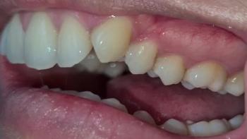
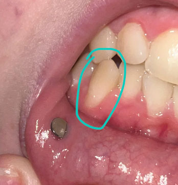
The circled tooth shows localized gum recession on a lower premolar, with visible root surface exposure. The gum margin has migrated downward, exposing the lighter-colored root area compared to the enamel above. The surrounding gum tissue appears mildly inflamed but without obvious pus or acute infection.
A metal stud visible on the lip is unrelated to the dental condition.
This presentation is consistent with early-to-moderate gingival recession, not active tooth decay.
Primary Diagnosis
-
Localized gingival recession on a lower premolar
-
Exposed root surface (cementum/dentin exposure)
-
Early non-carious cervical lesion risk
-
Increased susceptibility to tooth sensitivity and root caries
Deep Clinical Examination (Image-Based Findings)
-
Gum margin positioned apical to its normal level
-
Smooth, light-colored exposed root surface
-
Mild gingival inflammation at the recession margin
-
No visible abscess, ulceration, or necrotic tissue
-
Likely contributing factors:
-
Aggressive or improper brushing technique
-
Thin gum biotype
-
Local plaque accumulation
-
Occlusal stress or tooth position
-
Can This Heal in 14 Days?
Gum recession does not reverse naturally.
However, within 14 days, appropriate dental care can:
-
Stop further recession
-
Reduce inflammation
-
Decrease or prevent sensitivity
-
Protect the exposed root surface
Early management is key to long-term stability.
Recommended Treatment Process
Step 1: Periodontal Assessment (Days 1–3)
-
Periodontal probing to measure recession depth
-
Evaluation of gum thickness and attachment level
-
Assessment of brushing habits and bite forces
Step 2: Disease Control and Prevention (Days 3–7)
-
Professional scaling and polishing
-
Removal of plaque near the gum line
-
Instruction on gentle brushing technique
-
Use of desensitizing toothpaste if sensitivity exists
Step 3: Protective or Corrective Care (Days 7–14)
Depending on severity and symptoms:
-
Fluoride or desensitizing varnish
-
Cervical composite restoration if root wear progresses
-
Occlusal adjustment if traumatic bite is present
-
Periodontal referral if recession continues to worsen
Expected Healing Timeline
-
Reduction in gum inflammation: 7–10 days
-
Sensitivity improvement: 1–2 weeks
-
Long-term stabilization depends on habit correction and maintenance
Note: Gum tissue will stabilize but will not regrow without surgical intervention.
What Will Escalate If Untreated
-
Increased tooth sensitivity
-
Higher risk of root caries
-
Progressive gum recession
-
Aesthetic concerns due to elongated tooth appearance
-
Potential future tooth mobility in advanced cases
Professional Comments
This is a common and manageable dental condition. When addressed early, most patients maintain their teeth long-term without surgery. The key is early prevention and habit correction, not aggressive treatment.
This is not an emergency, but it should not be ignored.
Strong Recommendation
Schedule a dental visit for periodontal evaluation and preventive treatment.
You can find a dental clinic near your area using this directory:
https://cebudentalimplants.com/map-dental-clinic
Early care prevents more complex and costly procedures later.
Related Clinical Cases
Patients with lower front gum inflammation may also experience tartar buildup, periodontal disease, gum abscesses, or tooth decay. Explore these related clinical case studies.