Dental Case: Problems

Root Canal Infection With Apical Bone Loss - Dental Case Analysis
Severity: 
Teeth Problems: 

Dental Case Analysis – Root Canal Infection With Apical Bone Loss

Case Overview

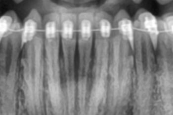
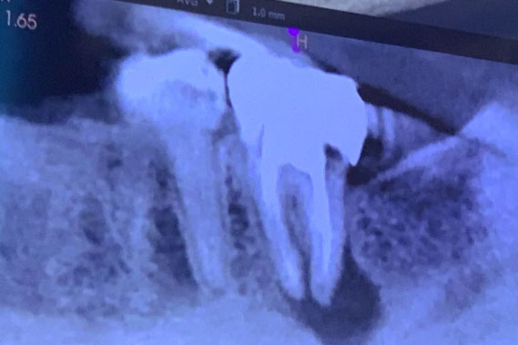
This dental X-ray shows a posterior tooth with previous root canal treatment. A clear radiolucent area at the root apex is visible, indicating a persistent or recurrent infection. The surrounding bone shows signs of loss consistent with chronic inflammation.


Full Analysis and Diagnosis

Radiographic Findings

  • Root canal–treated tooth with dense coronal restoration

  • Apical radiolucency at the root tip
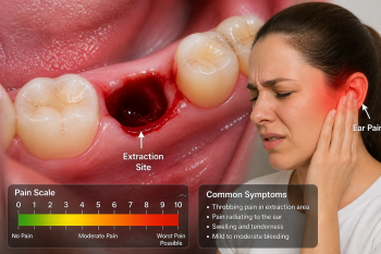
The Hidden Threat: What Back Tooth Pain and Bleeding Gums Could Mean for Your Health

Severity: 

Deep Examination & Analysis

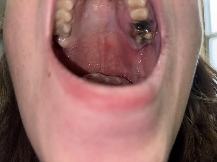
Based on the image provided, there are several significant dental health concerns that require immediate professional attention:

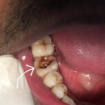
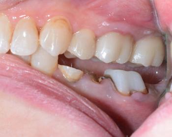
  • Gross Caries (Severe Decay): The lower molar (bottom right of the photo) shows a large, dark cavitation covering most of the occlusal (biting) surface. This indicates extensive tooth structure loss.

  • Likely Pulp Exposure: Given the depth of the dark area on the lower molar, the decay has likely reached the pulp (nerve) of the tooth.

Post-Extraction Swelling & Bruising - Is It Dry Socket or Normal Healing?

Severity: 

Quick summary of the situation you described

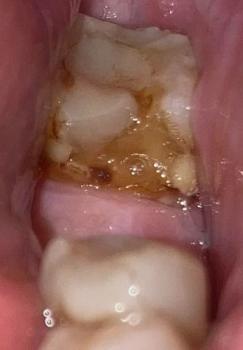
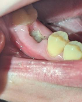
  • Tooth was extracted 2 days ago.

  • Swelling started before extraction and has not gone down since.

  • Area is now bruised. Extraction was difficult/traumatic.

  • You did not take antibiotics (previous C. difficile).

  • You cannot take typical OTC pain meds (alpha-gal + stomach issues).

Apical Infection After Root Canal - CBCT Dental Case

Severity: 

Dental Case Analysis – Apical Infection After Root Canal Treatment

Case Overview

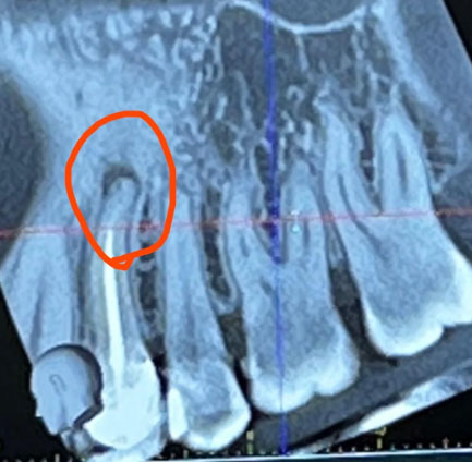
This CBCT image shows the anterior maxillary region with multiple natural teeth. A well-defined radiolucent lesion is visible at the root apex of a previously treated tooth (circled area), indicating a persistent apical infection. Surrounding bone shows early structural changes.


Full Analysis and Diagnosis

Radiographic Findings

  • Clear apical radiolucency at root tip