Missing Lower Molar With Braces Case Analysis
Severity:
Teeth Problems:
Missing Lower Molar With Braces Case Analysis and Care Plan
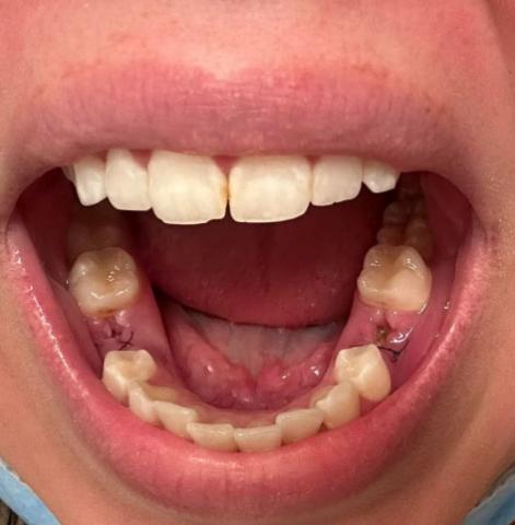
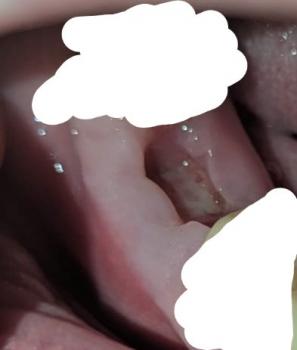
What Is Seen in This Case
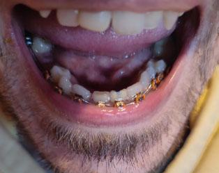
The image shows lower teeth with orthodontic braces, and a missing lower molar creating an open space along the dental arch. Brackets and wires are present on the remaining teeth, and plaque buildup is visible around orthodontic components. The gum tissue near the missing tooth appears healed but vulnerable.
This situation increases the risk of food trapping, tooth movement, and uneven bite forces during orthodontic treatment.