Dentures That Feel Natural, Look Beautiful.

Severe Decay of a Lower Molar: Expert Dental Assessment, Tooth-Saving Options, and Patient FAQs
Image:
Severity:
Teeth Problems:
Expert Dental Commentary: Why Severe Lower Molar Decay Requires Prompt Treatment
A lower molar plays an important role in chewing and maintaining normal bite function. When decay progresses deeply into the tooth, it can destroy enamel and dentin, weaken the remaining tooth structure, and eventually affect the dental pulp—the soft tissue containing nerves and blood vessels.
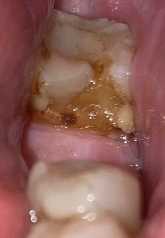
In the clinical case presented in this article, the lower molar shows substantial brown discoloration and visible breakdown of the chewing surface. These findings are concerning for advanced dental caries. However, a photograph alone cannot determine the exact depth of decay, whether the pulp is infected, or whether the tooth can still be restored.
The most important clinical question is whether enough healthy tooth structure remains to save the molar.
A dentist will evaluate the tooth's appearance, symptoms, response to pulp tests, gum condition, and dental X-rays before recommending treatment. The assessment may also include checking for cracks, tenderness when biting, and changes around the tooth roots.
Can a Severely Decayed Lower Molar Still Be Saved?
Yes, some severely decayed molars can still be preserved. The appropriate treatment depends on how much tooth structure remains and whether the pulp and surrounding tissues are affected.
-
Dental filling: Suitable when the decay can be removed and enough healthy tooth structure remains to support a restoration.
-
Dental crown: May be recommended when extensive damage has weakened the remaining tooth and it needs additional protection.
-
Root canal treatment: May be needed when the pulp is irreversibly inflamed or infected. The canals are cleaned, disinfected, and sealed before the tooth is restored.
-
Extraction: May be necessary when decay extends too far below the gumline, the tooth is severely fractured, or insufficient healthy structure remains for predictable restoration.
A root canal does not automatically mean the tooth is beyond repair. In appropriate cases, it can preserve the natural tooth. However, the tooth usually needs a suitable final restoration to protect it against fracture.
Why Waiting Can Make the Problem Worse
Early tooth decay may cause little or no discomfort. As the lesion progresses, patients may develop sensitivity to sweets, cold or hot drinks, pain while chewing, or persistent toothache.
If bacteria reach the pulp and infection develops around the root, an abscess may form. The infection can cause localized swelling, pus, tenderness, or fever. Some dental infections can spread into the face and neck.
Importantly, the absence of pain does not prove that the tooth is healthy. In some cases, the pulp may lose its vitality, reducing pain even while disease continues.
Patients with visible deep decay should arrange a dental examination promptly rather than waiting for severe pain to begin.
Short Patient Story: The Molar That Looked Like a Small Problem
Illustrative educational story—not a verified patient testimonial.
A patient noticed a dark brown area on a lower back tooth. At first, the discoloration seemed harmless, and occasional sensitivity disappeared after eating. Because there was no continuous toothache, the patient postponed a dental appointment.
Several weeks later, chewing on that side became uncomfortable. The patient finally sought professional care, hoping a simple filling would solve the problem.
During the examination, the dentist explained that the visible dark area could not reveal the full extent of the damage. An X-ray and additional tests were needed to assess the tooth's internal condition and remaining structure.
The dentist discussed the available options, including whether the molar could be preserved with restorative treatment or required a more extensive procedure.
The key lesson was simple: a tooth can be seriously damaged before the pain becomes severe. Early assessment gives patients a better opportunity to understand their options before the condition progresses.
What Happens During a Professional Dental Assessment?
A typical evaluation of severe lower molar decay includes the following steps:
-
Visual examination: The dentist checks the size and location of the cavity, remaining tooth structure, gum condition, and possible fractures.
-
Dental X-ray: Imaging helps assess decay depth, the roots, and the surrounding bone. The type of X-ray depends on the clinical findings.
-
Pulp assessment: Appropriate sensitivity or vitality tests help determine whether the nerve tissue is healthy, inflamed, or possibly non-vital.
-
Restorability assessment: The dentist evaluates whether the remaining tooth can support a durable restoration and whether the surrounding tissues are healthy enough for treatment.
-
Individualized treatment planning: The dentist explains the benefits, limitations, expected follow-up, and costs of the available options.
Not every patient needs a root canal, and not every severely decayed tooth needs extraction. Treatment should be based on the clinical findings rather than appearance alone.
Home Care Before the Dental Appointment
Home care can help control discomfort temporarily, but it cannot rebuild a tooth that has lost substantial structure or eliminate an established infection inside the tooth.
While waiting for professional treatment:
-
Brush gently twice daily using fluoride toothpaste.
-
Clean between the teeth carefully to reduce plaque accumulation.
-
Choose soft foods and chew on the opposite side if chewing is uncomfortable.
-
Avoid foods and drinks that trigger sensitivity, especially very hot, cold, or sugary items.
-
Consider gentle warm salt-water rinsing for temporary comfort, then spit it out.
-
Use an over-the-counter pain reliever only if it is safe for you and according to its label or a pharmacist's advice.
Do not place aspirin directly on the tooth or gum, attempt to dig out the decay, or use leftover antibiotics without professional guidance.
Antibiotics are not a substitute for treating the affected tooth and are not routinely indicated for most cases of pulpal dental pain. A dentist determines whether antibiotics are appropriate, particularly if there are signs of systemic illness or spreading infection.
When Is Severe Molar Decay a Dental Emergency?
Seek urgent dental care if you develop severe or persistent toothache, pus, increasing gum or facial swelling, fever, or difficulty opening your mouth.
Go to an emergency department immediately if swelling affects breathing or swallowing, spreads significantly into the neck, or is accompanied by other signs of a potentially life-threatening infection. Do not wait for a routine dental appointment in these circumstances.
Prompt treatment can reduce the risk of complications and help determine whether the natural molar can be preserved.
Frequently Asked Questions About Severe Lower Molar Decay
1. Can a severely decayed lower molar be saved without extraction?
Sometimes. A dentist may recommend a filling, a crown, root canal treatment followed by restoration, or a combination of procedures. Extraction is considered when the tooth cannot be restored predictably or has other serious problems.
2. Does brown or black discoloration always mean severe tooth decay?
No. Discoloration can result from decay, staining, or other changes in the tooth. A cavity, soft tooth structure, or breakdown of the chewing surface raises concern, but the diagnosis requires a dental examination.
3. Can severe tooth decay heal naturally?
A cavity that has already formed cannot reliably repair itself. Very early mineral loss before a cavity develops may sometimes be arrested or remineralized with fluoride and effective preventive care. Established structural damage usually requires professional treatment.
4. Will a severely decayed molar always need a root canal?
No. Root canal treatment is considered when the pulp is irreversibly inflamed or infected, or when other clinical findings indicate it is necessary. If the pulp remains healthy and the tooth is restorable, a filling or other restoration may be sufficient.
5. How do I know if decay has reached the tooth nerve?
Possible symptoms include prolonged sensitivity to hot or cold, spontaneous toothache, throbbing pain, or pain that disturbs sleep. However, symptoms alone cannot confirm the pulp's condition. Dental examination and appropriate tests are necessary.
6. Can a tooth infection exist without pain?
Yes. A non-vital tooth or a chronic infection may cause limited symptoms. A lack of pain does not exclude significant decay, root infection, or surrounding tissue damage.
7. Can antibiotics cure a decayed lower molar?
No. Antibiotics do not remove decay or restore damaged tooth structure. Most cases require treatment of the source, such as removing decay, root canal treatment, drainage when indicated, or extraction. Antibiotics may be appropriate in selected cases of spreading infection or systemic involvement.
8. Is extraction better than root canal treatment for a severely damaged molar?
Neither option is universally better. If the tooth can be restored successfully, preserving it may maintain natural chewing function. If the damage is too extensive, extraction may be the safer and more predictable choice. Your dentist should explain the prognosis and alternatives.
9. What happens if I leave severe lower molar decay untreated?
The decay may enlarge, weaken the tooth, cause fracture, reach the pulp, or lead to infection and abscess formation. Treatment may become more complex, and the tooth may eventually become impossible to save.
10. How can I prevent another molar from developing severe decay?
Brush twice daily with fluoride toothpaste, clean between your teeth daily, reduce frequent consumption of sugary foods and drinks, and attend regular dental check-ups. Your dentist may recommend additional fluoride or preventive measures based on your individual risk.
Final Expert Takeaway
Severe lower molar decay should not be judged by color or pain alone. The most reliable way to determine the next step is a professional examination supported by appropriate diagnostic tests and X-rays.
Early assessment may allow a tooth-preserving treatment plan. If the tooth cannot be saved, your dentist can discuss extraction and suitable replacement options when appropriate.
Do not wait for facial swelling or unbearable toothache before seeking care. Timely treatment protects your oral health and reduces the risk of a dental problem becoming an emergency.
Medical information notice: This article is for general educational purposes and does not replace an individual examination, diagnosis, or treatment plan from a licensed dental professional.
Severe Decay on Lower Molar Case Analysis and Treatment Plan
What Is Seen in This Case
The image shows a lower back tooth (molar) with heavy brown discoloration and visible decay on the chewing surface. The tooth structure appears weakened, with plaque, food debris, and possible softening of enamel. The surrounding gum tissue looks irritated, suggesting ongoing bacterial activity.
This is a clear sign of advanced tooth decay that has been developing over time.
Most Likely Diagnosis
Based on visual examination, the most likely conditions include:
-
Advanced dental caries on a lower molar
-
Breakdown of tooth enamel and dentin
-
High plaque and bacterial accumulation
-
Risk of pulp (nerve) involvement
-
Early gum inflammation around the tooth
A dental X-ray is required to confirm how deep the decay has reached.
What Causes This Level of Tooth Decay
Common causes include:
-
Long-term plaque buildup
-
Frequent sugary or acidic food intake
-
Inadequate brushing of back teeth
-
Delayed dental visits
-
Dry mouth or poor saliva flow
Lower molars are especially prone to decay because they are harder to clean.
Is This a Serious Problem
Yes. This condition is serious and active. If left untreated, it will scale up into:
-
Severe tooth pain
-
Tooth nerve infection
-
Dental abscess formation
-
Swelling of gums or face
-
Tooth fracture
-
Tooth loss
At this stage, the tooth cannot heal on its own.
Recommended Treatment Process
Initial Assessment (Days 1–3)
-
Full dental examination
-
Dental X-ray to assess decay depth
-
Evaluation of tooth restorability
Active Treatment Phase (Days 4–7)
Depending on severity, treatment may include:
-
Deep cleaning and removal of decayed material
-
Dental filling if the nerve is not involved
-
Root canal treatment if the nerve is infected
-
Temporary medication if infection is present
Final Restoration or Healing Phase (Days 8–14)
-
Permanent filling or crown placement
-
Monitoring of gum healing
-
Pain and sensitivity should reduce
If the tooth is too damaged, extraction may be recommended.
Expected Healing Time
-
Mild decay after filling: 3–7 days
-
Root canal–treated tooth: 7–14 days for comfort
-
Gum irritation improvement: within 14 days
Healing depends on how early treatment begins.
What Happens If Treatment Is Delayed
If delayed beyond 14 days, the condition may worsen and lead to:
-
Sudden severe toothache
-
Spreading infection
-
Abscess and pus formation
-
Emergency dental treatment
-
Higher treatment cost
-
Tooth loss
Early care prevents emergencies.
Home Care While Waiting for Treatment
These steps may reduce discomfort but do not stop decay:
-
Avoid chewing on the affected side
-
Rinse gently with warm salt water
-
Avoid sugary and sticky foods
-
Brush gently but thoroughly
Seek dental care as soon as possible.
Professional Comment
This case shows severe decay on a lower molar, already at high risk of nerve infection. Prompt dental treatment is critical to save the tooth and prevent abscess or extraction.
Visit a Dental Clinic Near You
For urgent dental evaluation and treatment, visit:
https://cebudentalimplants.com/map-dental-clinic
Related Clinical Cases
Patients with lower front gum inflammation may also experience tartar buildup, periodontal disease, gum abscesses, or tooth decay. Explore these related clinical case studies.




Other topics:
Other Dental Clinic
 |
 |
 |
 |