Because Every Tooth Deserves Care.
Severe Decay on Lower Molar Case Analysis
Image:
Severity:
Teeth Problems:
Severe Decay on Lower Molar Case Analysis and Treatment Plan
What Is Seen in This Case
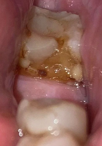
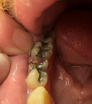
The image shows a lower back tooth (molar) with heavy brown discoloration and visible decay on the chewing surface. The tooth structure appears weakened, with plaque, food debris, and possible softening of enamel. The surrounding gum tissue looks irritated, suggesting ongoing bacterial activity.
This is a clear sign of advanced tooth decay that has been developing over time.
Most Likely Diagnosis
Based on visual examination, the most likely conditions include:
-
Advanced dental caries on a lower molar
-
Breakdown of tooth enamel and dentin
-
High plaque and bacterial accumulation
-
Risk of pulp (nerve) involvement
-
Early gum inflammation around the tooth
A dental X-ray is required to confirm how deep the decay has reached.
What Causes This Level of Tooth Decay
Common causes include:
-
Long-term plaque buildup
-
Frequent sugary or acidic food intake
-
Inadequate brushing of back teeth
-
Delayed dental visits
-
Dry mouth or poor saliva flow
Lower molars are especially prone to decay because they are harder to clean.
Is This a Serious Problem
Yes. This condition is serious and active. If left untreated, it will scale up into:
-
Severe tooth pain
-
Tooth nerve infection
-
Dental abscess formation
-
Swelling of gums or face
-
Tooth fracture
-
Tooth loss
At this stage, the tooth cannot heal on its own.
Recommended Treatment Process
Initial Assessment (Days 1–3)
-
Full dental examination
-
Dental X-ray to assess decay depth
-
Evaluation of tooth restorability
Active Treatment Phase (Days 4–7)
Depending on severity, treatment may include:
-
Deep cleaning and removal of decayed material
-
Dental filling if the nerve is not involved
-
Root canal treatment if the nerve is infected
-
Temporary medication if infection is present
Final Restoration or Healing Phase (Days 8–14)
-
Permanent filling or crown placement
-
Monitoring of gum healing
-
Pain and sensitivity should reduce
If the tooth is too damaged, extraction may be recommended.
Expected Healing Time
-
Mild decay after filling: 3–7 days
-
Root canal–treated tooth: 7–14 days for comfort
-
Gum irritation improvement: within 14 days
Healing depends on how early treatment begins.
What Happens If Treatment Is Delayed
If delayed beyond 14 days, the condition may worsen and lead to:
-
Sudden severe toothache
-
Spreading infection
-
Abscess and pus formation
-
Emergency dental treatment
-
Higher treatment cost
-
Tooth loss
Early care prevents emergencies.
Home Care While Waiting for Treatment
These steps may reduce discomfort but do not stop decay:
-
Avoid chewing on the affected side
-
Rinse gently with warm salt water
-
Avoid sugary and sticky foods
-
Brush gently but thoroughly
Seek dental care as soon as possible.
Professional Comment
This case shows severe decay on a lower molar, already at high risk of nerve infection. Prompt dental treatment is critical to save the tooth and prevent abscess or extraction.
Visit a Dental Clinic Near You
For urgent dental evaluation and treatment, visit:
https://cebudentalimplants.com/map-dental-clinic
Teeth Case
 |
 |
 |
 |
 |
 |