Modern Dentistry, Timeless Smiles.
Exposed Gum Tissue Near Lower Molar Case Analysis
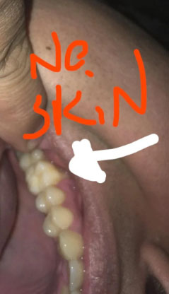
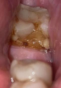
Image:
Severity:
Teeth Problems:
Exposed Gum Tissue Near Lower Molar Case Analysis (No Protective Skin)
What Is Seen in This Case
The image shows an area near the lower back teeth where the gum tissue appears thin or missing, exposing the underlying soft tissue. The marked area (“no skin”) suggests loss of normal gum coverage, which can make the area sensitive, sore, and vulnerable to infection.
The nearby teeth appear intact, but the gum condition indicates localized tissue damage or recession.
Most Likely Diagnosis
Based on visual examination, the most likely conditions include:
-
Gum recession near the lower molar
-
Loss of protective gum tissue
-
Localized gum trauma or irritation
-
Early infection risk due to exposed tissue
-
Possible post-extraction or post-inflammation tissue loss
A dental examination is required to determine the cause and depth of tissue involvement.
What Causes Loss of Gum “Skin” in This Area
Common causes include:
-
Aggressive brushing or repeated friction
-
Previous tooth extraction or dental surgery
-
Chronic gum inflammation
-
Food impaction causing repeated irritation
-
Poor oral hygiene leading to tissue breakdown
Once gum tissue is damaged, it does not easily regrow without care.
Is This a Serious Problem
This condition can become serious if ignored. If left untreated, it may scale up into:
-
Persistent pain or sensitivity
-
Infection of the exposed tissue
-
Worsening gum recession
-
Bone exposure in advanced cases
-
Increased risk of tooth loss
-
Difficulty chewing comfortably
Early treatment helps protect the area and prevent progression.
Recommended Treatment Process
Initial Assessment (Days 1–3)
-
Dental examination
-
Evaluation of gum thickness and health
-
Check for infection or trauma source
Active Treatment Phase (Days 4–7)
Treatment may include:
-
Professional cleaning
-
Medication or topical treatment if inflamed
-
Protection of the exposed area
-
Adjustment of brushing technique
Healing and Protection Phase (Days 8–14)
-
Gum tissue irritation should reduce
-
Sensitivity should improve
-
Further treatment planned if tissue loss persists
In some cases, gum grafting or specialist care may be recommended.
Expected Healing Time
-
Mild tissue irritation: 5–7 days
-
Moderate gum damage: up to 14 days
-
Long-term tissue repair depends on severity and treatment
What Happens If Treatment Is Delayed
If delayed beyond 14 days, this condition may worsen and cause:
-
Ongoing pain and sensitivity
-
Gum infection
-
Progressive tissue loss
-
Bone exposure
-
Higher treatment cost
Gum tissue problems should be addressed early.
Home Care While Waiting for Treatment
These steps help reduce irritation but do not replace dental care:
-
Brush gently with a soft toothbrush
-
Avoid touching the area with fingers or tongue
-
Rinse gently with warm salt water
-
Avoid spicy, hot, or hard foods
Professional Comment
This case shows loss of protective gum tissue near a lower molar, making the area vulnerable. Early dental evaluation is important to prevent infection, protect bone, and restore comfort.
Visit a Dental Clinic Near You
For gum evaluation and treatment planning, visit:
https://cebudentalimplants.com/map-dental-clinic
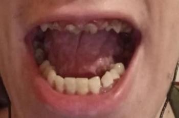
Teeth Case
 |
 |
 |
 |
 |
 |