Healthy Teeth, Happy Life.

Dental Hub Clinical Case Study: Dentigerous Cyst
A dentigerous cyst is a developmental odontogenic cyst that typically develops around the crown of an unerupted or impacted tooth. It is frequently discovered incidentally because many patients have no symptoms during the early stages. Radiographically, it classically appears as a well-defined radiolucent area surrounding the crown of an unerupted tooth.
Important: The following California case is presented as an educational clinical scenario, not as an identifiable real patient's medical record.
1. California Patient Presentation
Patient: Adult patient in California
Reason for dental evaluation: Routine dental examination and panoramic imaging
Initial symptoms: No significant pain; no obvious facial swelling
Radiographic finding: A well-defined radiolucent lesion surrounding the crown of an impacted mandibular third molar
The patient was surprised because there had been little indication that a significant lesion was developing.
This is a classic clinical characteristic of dentigerous cysts: they can remain silent while gradually expanding within the jawbone.
The clinical question
The finding raised an important question:
Was this simply an enlarged dental follicle, or was there a true cystic lesion—and could another odontogenic lesion be producing the same radiographic appearance?
2. Understanding the Dentigerous Cyst
A dentigerous cyst develops in association with the crown of an unerupted or impacted tooth.
Commonly involved teeth include:
-
Mandibular third molars
-
Maxillary canines
-
Maxillary third molars
-
Other unerupted permanent teeth
The mandibular third-molar region is particularly common.
On imaging, the characteristic pattern is a unilocular radiolucency surrounding the crown and extending toward the cemento-enamel junction.
However, radiographic appearance alone does not establish the final diagnosis.
3. Why This California Case Matters
The patient had no major symptoms.
That does not mean the lesion was clinically irrelevant.
As dentigerous cysts enlarge, they can cause:
-
Expansion of the jawbone
-
Facial asymmetry
-
Displacement of adjacent teeth
-
Root resorption
-
Bone thinning
-
Infection
-
Pressure on nearby anatomical structures
Large lesions can therefore transform a silent radiographic finding into a significant surgical problem.
From a U.S. dentist's perspective, this is one reason routine dental examinations and appropriately indicated radiographic evaluation remain important.
4. Diagnostic Work-Up
Clinical examination
The dentist evaluates:
-
Facial symmetry
-
Intraoral swelling
-
Tooth eruption status
-
Tooth mobility
-
Periodontal condition
-
Adjacent teeth
-
Neurologic symptoms
-
Signs of infection
In this hypothetical California case, the absence of significant pain or swelling did not eliminate the need for further evaluation.
Panoramic radiograph
The panoramic image demonstrated a well-circumscribed radiolucency surrounding the crown of the impacted tooth.
This appearance strongly suggested a dentigerous cyst.
But a responsible diagnosis requires consideration of other possibilities.
5. Differential Diagnosis
A lesion around an impacted tooth can potentially resemble several conditions.
The differential diagnosis may include:
-
Enlarged dental follicle
-
Odontogenic keratocyst
-
Unicystic ameloblastoma
-
Other odontogenic cysts
-
Other odontogenic tumors
Published pathology reviews specifically emphasize the importance of correlating the radiographic relationship to the affected tooth with the clinical and microscopic findings.
Clinical lesson
A radiographic "cyst-like" appearance is not automatically the final diagnosis.
6. CBCT: Looking at the Lesion in Three Dimensions
When additional anatomical information is clinically indicated, CBCT can provide a three-dimensional assessment.
The oral surgeon may want to determine:
-
Exact lesion size
-
Buccal and lingual cortical expansion
-
Cortical thinning or perforation
-
Relationship to adjacent tooth roots
-
Position of the impacted tooth
-
Relationship to the inferior alveolar canal
-
Surgical access
-
Extent of surrounding bone involvement
This information can be particularly valuable when planning surgery for a large mandibular lesion.
7. Treatment Planning
The appropriate treatment depends on several factors:
-
Patient age
-
Lesion size
-
Tooth involved
-
Position of the impacted tooth
-
Ability to preserve the tooth
-
Risk to adjacent structures
-
Amount of bone destruction
-
Clinical symptoms
-
Histopathological findings
There is no single treatment that is appropriate for every dentigerous cyst.
8. Enucleation
For many dentigerous cysts, treatment involves surgical removal of the cystic tissue, frequently together with removal of the associated impacted tooth when preservation is not considered predictable.
The cystic tissue may be carefully separated from the surrounding bone.
The surgical specimen should be handled appropriately for pathological examination when indicated.
Published reviews describe enucleation with management of the associated tooth as a common treatment approach, with conservative alternatives considered in selected cases.
9. Marsupialization and Decompression
A more conservative approach may be considered for selected large lesions, particularly when preservation of an unerupted permanent tooth is desirable.
Marsupialization creates a surgical opening into the cyst, allowing decompression and gradual reduction of the lesion.
This can potentially:
-
Reduce cyst size
-
Encourage bone formation
-
Protect nearby structures
-
Create an opportunity for an unerupted tooth to erupt
-
Reduce the extent of subsequent surgery
Case literature demonstrates that conservative management can allow an associated permanent tooth to erupt in appropriately selected patients.
10. Histopathology: The Final Confirmation
After surgical removal or biopsy, the tissue can be examined microscopically.
Classic dentigerous cysts demonstrate a connective-tissue wall with an epithelial lining, although inflammation can substantially alter the microscopic appearance.
Histopathology is important because lesions that look similar on an X-ray may have very different biological behavior.
For this reason, the clinical diagnosis should be correlated with the pathology report whenever tissue is obtained.
11. What Could Happen If the Cyst Is Ignored?
A small asymptomatic lesion may remain clinically quiet, but continued enlargement can create progressively more difficult problems.
Potential consequences include:
Bone expansion → cortical thinning → tooth displacement → root resorption → structural weakening
In severe cases, an extensive lesion can occupy a substantial portion of the jaw.
Recent case literature emphasizes that dentigerous cysts can remain clinically silent while producing considerable radiographic expansion.
12. California Case Outcome — Educational Scenario
For this hypothetical California patient, the clinical team would establish the lesion's size and anatomical relationships, evaluate the impacted tooth, and determine whether conservative management or surgical removal offered the most predictable result.
If the tooth could not reasonably be preserved, removal of the impacted tooth together with cyst enucleation would be considered.
If preservation and eruption were realistic—particularly in a younger patient—decompression or marsupialization could be considered.
The removed tissue would be evaluated according to the treating team's pathology protocol.
Follow-up imaging would then be used to monitor bone healing and confirm that the lesion has not returned.
13. U.S. Dental Expert Perspective
As a U.S. dental clinician, I would emphasize one important message to patients:
Do not judge a jaw lesion by symptoms alone.
A patient can feel perfectly healthy while an odontogenic cyst is developing around an impacted tooth.
The combination of:
Clinical examination + appropriate radiography + CBCT when indicated + pathological correlation
provides a much stronger basis for diagnosis and treatment planning than any single test.
14. Questions California Patients Commonly Ask
"Is a dentigerous cyst cancer?"
Usually, a dentigerous cyst is a benign developmental odontogenic cyst. However, the diagnosis should be established appropriately because other lesions can mimic its appearance.
"Does every dentigerous cyst need surgery?"
Not necessarily the same type of surgery. Management depends on the lesion and patient. Options can include enucleation or conservative decompression/marsupialization in selected cases.
"Can the impacted tooth be saved?"
Sometimes. In appropriately selected patients, particularly younger patients, decompression or marsupialization may allow an unerupted tooth to erupt or be orthodontically guided into position.
"Will it come back?"
Recurrence after appropriate treatment is generally uncommon, but long-term follow-up should be determined by the lesion's pathology, treatment, and clinical circumstances.
"Why did I have no pain?"
Because many dentigerous cysts are asymptomatic and discovered incidentally on dental imaging.
Clinical Takeaway
This California case illustrates an important principle in modern dental diagnosis:
A silent jaw lesion can still be clinically significant.
A dentigerous cyst may develop around an impacted tooth without producing pain or visible swelling. Once identified, the goal is to establish an accurate diagnosis, understand the lesion's three-dimensional anatomy, protect important structures, and select the least invasive treatment that provides a predictable long-term result.
For patients, the most important action is simple:
If your dentist discovers a cyst-like lesion around an impacted tooth, do not ignore it. Ask what the lesion represents, whether additional imaging is necessary, whether the tooth can be preserved, and whether an oral and maxillofacial surgeon or oral pathology evaluation is appropriate.
Dental Hub Disclaimer
This case is an educational California-based clinical scenario, not a report of an identifiable individual. It is not a substitute for an examination, diagnosis, or treatment plan from a licensed dental professional. Treatment decisions should be individualized according to the patient's clinical examination, imaging, pathology, medical history, and informed consent.
Related Clinical Cases
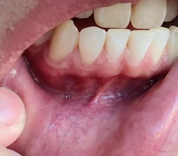
Patients with lower front gum inflammation may also experience tartar buildup, periodontal disease, gum abscesses, or tooth decay. Explore these related clinical case studies.


Other topics:
Other Dental Clinic