Healthy Teeth, Happy Life.

Severe Tartar Buildup on Lower Front Teeth: Gum Inflammation, Treatment, 14-Day Healing Timeline & Risk of Progression
Image:
Severity:
Teeth Problems:
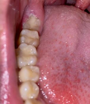
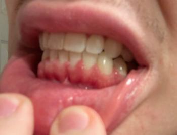
This is a clinical photo rather than an X-ray, so I can assess visible plaque/calculus, gum appearance, tooth alignment, and staining, but I cannot determine bone loss or periodontal pocket depth from the photo alone.
Main finding
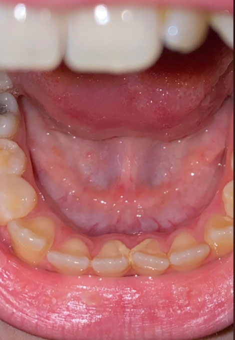
The most obvious issue is substantial calculus (tartar) accumulation on the lower front teeth, particularly around the lingual/cervical surfaces. There is also visible gingival redness/inflammation.
The lower incisors appear crowded/rotated, which can make plaque and calculus much easier to accumulate.
Estimated visual severity
| Finding | Visual assessment |
|---|---|
| Calculus/tartar | Moderate–severe |
| Plaque accumulation | Moderate |
| Gingival inflammation | Moderate |
| Lower anterior crowding | Moderate |
| Visible staining | Mild–moderate |
| Obvious pus/abscess | Not visible |
| Obvious severe ulceration | Not visible |
| Bone loss | Cannot determine from photo |
| Periodontitis | Cannot diagnose from photo alone |
What concerns me most
The calculus is not simply a cosmetic problem. Once hardened, calculus cannot be reliably removed with normal brushing. It provides a rough surface that retains bacterial plaque and can contribute to persistent gingival inflammation.
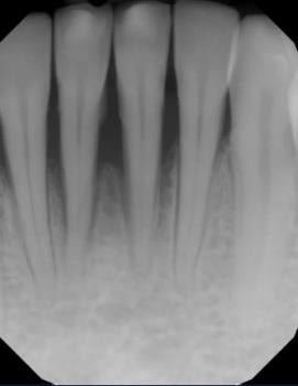
If the inflammation has been present for a long time, the next question is whether there is periodontal pocketing or bone loss underneath the gum. That requires periodontal probing and usually dental radiographs.
What should happen next?
A dentist should ideally perform:
- Comprehensive dental examination
- Periodontal probing around the affected teeth
- Professional scaling/polishing
- Evaluation for bleeding on probing
- Check for gum recession and tooth mobility
- Dental X-rays if periodontal disease is suspected
- Determine whether routine scaling is sufficient or whether deep periodontal cleaning/scaling and root planing is necessary
- Re-evaluation after healing
14-day timeline
If this is primarily gingivitis caused by plaque/calculus and the calculus is professionally removed:
Day 0: examination + professional cleaning
Days 1–3: gums may remain tender or bleed somewhat
Days 4–7: inflammation should begin noticeably decreasing
Days 7–14: gum tissue can become considerably healthier if plaque control is good
After 14 days: reassessment can determine whether inflammation has resolved or whether periodontal treatment is needed
Important: 14 days does not mean periodontal disease is completely cured. Soft-tissue inflammation can improve within about two weeks, while periodontal attachment/bone problems, if present, require much longer management.
Expert Guide: How to Fix Crooked, Crowded, and Gapped Teeth Safely
Can You Straighten Your Teeth in 14 Days? A Dentist Explains
Patients often ask whether crooked teeth, dental crowding, or gaps between teeth can be corrected within 14 days. The answer depends on what they mean by correction. While certain cosmetic procedures can change the appearance of teeth quickly, safely moving natural teeth into new positions requires biological changes in the surrounding bone and supporting tissues.
True orthodontic tooth movement cannot usually be completed in 14 days. Braces and clear aligners apply controlled forces that gradually move teeth through the supporting bone. The bone must remodel around the roots as the teeth change position. Attempting to accelerate this process excessively can increase the risk of root damage, gum recession, and other complications.
The appropriate treatment depends on the patient's bite, the severity of misalignment, gum health, tooth condition, and treatment goals.
1. How Dentists Correct Different Types of Tooth Misalignment
Not every alignment problem requires the same treatment. A comprehensive dental examination helps determine whether the problem involves tooth position, tooth shape, missing teeth, or the relationship between the upper and lower jaws.
Crooked teeth
Crooked teeth may rotate, tilt, or overlap neighboring teeth. Mild cases may be suitable for clear aligners or limited orthodontic treatment, while more complicated cases may require comprehensive braces.
The objective is not simply to make the front teeth look straight. The dentist must also consider root position, the bite, gum health, and long-term stability.
Crowded teeth
Dental crowding occurs when there is insufficient space for the teeth to fit properly within the dental arch. It can make brushing and flossing more difficult and may contribute to plaque accumulation.
Depending on the severity, treatment may involve braces, clear aligners, or carefully planned space creation. Some patients require tooth reshaping between selected teeth, called interproximal reduction, while others may need extractions. These decisions require an individualized assessment; extraction is not automatically necessary for every crowded smile.
Gapped teeth
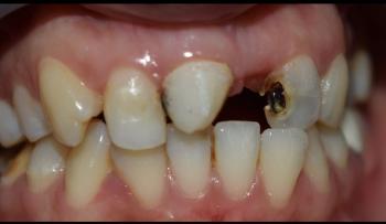
Spaces between teeth may occur because of naturally small teeth, missing teeth, differences in jaw size, habits, or periodontal disease.
Small gaps may sometimes be closed with composite bonding or orthodontic treatment. Larger spaces may require braces, clear aligners, veneers, crowns, or tooth replacement, depending on the underlying cause.
Important: New or widening gaps between teeth should not be treated as a cosmetic problem alone. Gum disease and loss of supporting bone can cause teeth to move apart. A dentist should investigate the cause before closing the spaces.
2. Braces vs. Clear Aligners vs. Cosmetic Bonding
The best option depends on whether the goal is to move teeth, change their appearance, or correct a functional problem.
| Treatment | Main purpose | Typical timeframe |
|---|---|---|
| Traditional braces | Move teeth and correct many types of bite problems | Often 18–24 months; some cases take longer |
| Clear aligners | Gradually reposition teeth using a series of custom trays | Commonly several months to a few years |
| Composite bonding | Change the visible shape or size of teeth | Often one appointment |
| Porcelain veneers | Change tooth appearance, including selected minor alignment concerns | Usually several appointments |
| Dental crowns | Restore teeth that are significantly damaged or weakened | Usually several appointments |
These timeframes are general estimates, not guarantees. Mild orthodontic cases may finish sooner, while complex crowding or bite problems can take substantially longer.
Cosmetic bonding and veneers do not physically straighten the tooth roots or correct the underlying bite. Veneers may require irreversible removal of some natural enamel, so they should not be used as a shortcut when a more conservative treatment is appropriate.
3. What Can Realistically Happen in the First 14 Days?
The first two weeks of orthodontic treatment are generally an adjustment period, not the completion stage.
During this period, patients may experience mild pressure or tenderness as the teeth begin responding to treatment. Some patients may notice small early changes, but visible progress varies considerably.
A realistic timeline looks like this:
-
Days 1–3: The patient adapts to braces or the first aligner trays. Mild soreness or pressure may occur.
-
Days 4–14: Initial tooth movement may be underway. The patient should follow the prescribed appliance-wearing schedule and maintain careful oral hygiene.
-
Following months: More noticeable alignment changes may develop, depending on the treatment plan and the complexity of the case.
-
After active treatment: Retainers help prevent the teeth from drifting toward their previous positions.
Patients should never attempt to speed up tooth movement by wearing aligners more aggressively than prescribed, forcing trays into place, or using homemade orthodontic devices.
4. Can Cosmetic Dentistry Make Teeth Look Straighter Quickly?
In selected cases, yes. Cosmetic dentistry can improve the appearance of minor irregularities more quickly than orthodontics, but it does not produce the same biological result.
For example, composite bonding may improve the appearance of a small gap or an uneven tooth edge. Veneers may alter the visible contours of selected teeth. However, these treatments are not suitable for every patient, especially when there is significant crowding, an abnormal bite, or active gum disease.
Before recommending a cosmetic procedure, the dentist should evaluate:
-
The health of the teeth and gums.
-
The amount of natural tooth structure that would need to be altered.
-
Whether the proposed change would interfere with the bite.
-
The likelihood of chipping, wear, or future repairs.
-
Whether orthodontic treatment could achieve a more conservative result.
A faster cosmetic result is not necessarily a better long-term treatment.
5. Why a Professional Orthodontic Examination Matters
Tooth alignment is a biological and functional process, not simply a matter of appearance. Before treatment begins, the dentist or orthodontist may assess the bite, tooth positions, gum condition, jaw relationship, and existing restorations.
Dental photographs, digital scans or impressions, and X-rays may be recommended when clinically indicated. These help the clinician develop an appropriate treatment plan and identify problems that may not be visible in a photograph.
Active gum disease, untreated tooth decay, and poor oral hygiene should be addressed before orthodontic treatment proceeds. Moving teeth when their supporting tissues are unhealthy can increase the risk of complications.
DIY braces, rubber bands placed around teeth, and unsupervised tooth-straightening kits can damage the gums and supporting bone, cause unwanted tooth movement, or contribute to tooth loss.
6. How to Maintain Healthy Teeth During Alignment Treatment
Good oral hygiene is essential throughout orthodontic treatment.
-
Brush twice daily with fluoride toothpaste.
-
Clean carefully around brackets and along the gumline.
-
Clean between teeth daily using floss or an appropriate interdental cleaner.
-
Limit frequent sugary snacks and drinks.
-
Attend scheduled dental and orthodontic appointments.
-
Wear removable aligners for the prescribed number of hours and remove them for meals and drinks other than water, as instructed.
-
Clean aligners according to the manufacturer's and clinician's instructions.
After treatment, wear the prescribed retainer. Teeth can shift over time even after successful orthodontic correction, so retention is an essential part of maintaining the result.
When Should You See a Dentist Promptly?
Arrange a dental assessment if your teeth are becoming loose, gaps are suddenly widening, your gums bleed persistently, or your bite changes unexpectedly. These symptoms can indicate gum disease or another underlying problem.
Severe pain, facial swelling, fever associated with dental symptoms, or difficulty opening the mouth warrants urgent professional assessment. Difficulty breathing or swallowing, or rapidly spreading swelling involving the face or neck, requires emergency care.
Expert Dentist's Final Advice
Straightening crooked, crowded, or gapped teeth can improve appearance and, in suitable cases, oral function and cleaning access. However, the safest treatment is one that addresses the underlying problem rather than simply producing a quick cosmetic change.
If your goal is to improve your smile within 14 days, ask a dentist whether a conservative cosmetic option is appropriate. If your goal is to move teeth into healthier positions, expect a treatment plan based on your individual anatomy and clinical needs.
The priority should be healthy teeth, stable gums, a functional bite, and a result that lasts—not the fastest possible treatment.
Frequently Asked Questions About Teeth Alignment
1. Can I fix crooked teeth in 14 days?
Usually not with orthodontics. Braces and clear aligners require time for the supporting bone to remodel as the teeth move. Some cosmetic procedures can improve the visible appearance of selected teeth in one or more visits, but they do not provide equivalent orthodontic correction.
2. What is the fastest way to straighten teeth?
The fastest appropriate option depends on the cause and severity of the problem. Mild alignment concerns may be treated with limited orthodontics or clear aligners. Cosmetic bonding can improve selected small irregularities without moving the teeth. A dental examination is necessary to determine the safest choice.
3. How long does it take to fix crowded teeth with braces?
Many comprehensive orthodontic treatments take approximately 18–24 months, although mild cases may take less time and complex cases may take longer. The estimate depends on crowding, bite correction, tooth movement, patient cooperation, and treatment response.
4. Are clear aligners better than traditional braces?
Neither option is universally better. Clear aligners are removable and discreet, but they require consistent wear and may not be suitable for every complex movement. Traditional braces can treat a wide range of alignment and bite problems. Your dentist or orthodontist can explain which method is appropriate for your case.
5. Can gaps between teeth close naturally?
Some spacing may change during growth, but adults should not rely on gaps to close naturally. New or increasing gaps can indicate gum disease, bone loss, missing teeth, or changes in tooth position. A dentist should determine the cause before treatment.
6. Can dental bonding fix gapped teeth in one day?
Small gaps may be improved with composite bonding during one appointment. However, bonding changes the visible shape of the teeth rather than moving them. It may not be appropriate for large gaps, significant bite problems, or gaps caused by active periodontal disease.
7. Do veneers straighten crooked teeth?
Veneers can change the appearance of selected mildly irregular teeth, but they do not reposition the roots or correct the underlying bite. Because veneers may require removal of natural enamel, the dentist should consider less invasive alternatives first.
8. Is teeth alignment painful?
Braces and aligners may cause temporary pressure or tenderness, particularly when treatment begins or an appliance is adjusted. The discomfort is usually manageable and tends to improve over several days. Severe or persistent pain should be assessed by the treating clinician.
9. Can adults straighten their teeth?
Yes. Healthy adult teeth can be moved orthodontically. Treatment planning must account for gum health, bone support, existing fillings or crowns, missing teeth, and any other dental conditions.
10. Can I use rubber bands to close a gap between my teeth?
No. Do not place ordinary rubber bands around teeth to close spaces. They can slip beneath the gumline, damage supporting tissues, and potentially cause tooth loss. Gap closure should be planned and monitored by a qualified dental professional.
11. Will my teeth become crooked again after braces?
Teeth can shift after orthodontic treatment. Retainers help maintain the result while the surrounding tissues adapt and over the long term. Follow your orthodontist's instructions for retainer wear and replacement.
12. Should I straighten my teeth if I have bleeding gums?
Bleeding gums should be evaluated before orthodontic treatment begins. Gingivitis and periodontitis require appropriate management, and active periodontal disease may need stabilization before tooth movement is considered safe.
13. Do I need to extract teeth to correct crowding?
Not always. Some cases can be treated by other methods of creating or managing space. Extractions may be appropriate when there is substantial crowding or a specific treatment objective, but the decision must be based on a comprehensive assessment.
14. Can teeth alignment improve oral health?
It can help some patients clean difficult-to-reach areas and may improve certain bite-related problems. However, straight teeth are not automatically healthy teeth. Daily plaque control, fluoride use, a balanced diet, and regular dental examinations remain essential.
15. What is the first step toward fixing crooked or gapped teeth?
Schedule an examination with a dentist or orthodontist. The clinician can identify the cause of the misalignment, evaluate gum and tooth health, discuss suitable treatment options, and provide a realistic timeline and cost estimate.
What could scale up if treatment is delayed?
If the patient waits 14 days, the calculus itself is unlikely to suddenly become dramatically worse. The bigger concern is continued bacterial plaque and inflammation.
If left untreated for months/years, the progression can become:
Plaque → calculus → gingivitis → periodontal pocket formation → attachment loss → bone loss → gum recession → tooth mobility → possible tooth loss.
So I would classify this as something that deserves dental treatment soon, but the photograph does not show an obvious emergency.
If there is spontaneous bleeding, bad breath, gum swelling, pus, loose teeth, significant pain, or rapidly increasing swelling, the evaluation should be accelerated.
Tartar, Gum Inflammation, Treatment & Healing
Important medical guidance: Tartar (dental calculus) is hardened plaque that cannot be safely removed completely with ordinary brushing. Professional dental cleaning is usually required. Severe tartar can contribute to gingivitis and, if periodontal disease develops, inflammation and destruction of the tissues and bone supporting the teeth.
What Severe Tartar Can Mean for Your Gums
Heavy tartar buildup around the lower front teeth is common because saliva from the nearby salivary glands can promote mineralization of plaque in this area. However, the amount of tartar alone does not determine how advanced gum disease is.
A dental professional should evaluate:
- Gum redness and swelling
- Bleeding during brushing or examination
- Gum pocket depths
- Gum recession
- Tooth mobility
- Plaque and calculus deposits below the gumline
- Bone loss on dental X-rays when clinically indicated
- Bad breath or persistent unpleasant taste
A person can have substantial tartar with mainly gingivitis, while another person with less visible tartar may already have periodontal disease.
Can Severe Tartar Cause Bone Loss?
Tartar itself does not directly "eat away" the bone. The concern is the chronic bacterial plaque and inflammatory response associated with periodontal disease.
When periodontitis develops and remains untreated, inflammation can damage the periodontal ligament and supporting alveolar bone. Progressive bone loss can eventually cause teeth to become loose.
This is why severe tartar should not simply be treated as a cosmetic problem.
What Treatment May Be Needed?
Treatment depends on the condition of the gums and supporting tissues.
1. Professional dental cleaning
For deposits mainly above the gumline, a dentist or dental hygienist may perform scaling and polishing.
2. Scaling and root planing
If calculus and bacterial deposits extend beneath the gumline and periodontal pockets are present, deeper periodontal cleaning may be recommended. This is sometimes called scaling and root planing (SRP) or nonsurgical periodontal therapy.
3. Periodontal reassessment
After initial treatment, the dentist may reassess gum bleeding, pocket depths, inflammation and plaque control.
4. Additional periodontal treatment
If significant periodontal disease remains, referral to a periodontist or additional treatment may be appropriate.
What Should You Expect During the First 14 Days?
A "14-day healing timeline" should be viewed as a general example, not a guaranteed healing schedule.
Days 1–3:
Gums may remain tender or bleed slightly, particularly when they were severely inflamed before treatment. Some patients experience temporary tooth sensitivity.
Days 4–7:
Inflammation and bleeding may begin to decrease when professional treatment is combined with effective daily plaque control.
Days 8–14:
The gums may look and feel healthier, although deeper periodontal inflammation can require considerably longer to stabilize.
Importantly, healthy gum tissue and periodontal stability cannot always be judged after only 14 days. Your dentist may need to monitor the gums over several weeks or longer.
How to Help the Gums Heal
After professional treatment:
- Brush twice daily with a soft-bristled toothbrush.
- Clean between the teeth every day using floss or an appropriate interdental cleaner.
- Follow your dentist's instructions regarding mouthwash.
- Do not attempt to scrape tartar away with sharp objects.
- Avoid smoking or tobacco products because they can interfere with periodontal health and healing.
- Keep scheduled dental follow-up appointments.
- Maintain regular professional cleanings based on your individual risk.
When Should You See a Dentist Quickly?
Seek prompt dental evaluation if you have:
- Facial or jaw swelling
- Pus around a tooth or gum
- Fever
- Severe or worsening dental pain
- Difficulty swallowing or breathing
- Significant tooth mobility
- Rapidly increasing swelling
Facial swelling combined with difficulty breathing or swallowing can be an emergency and requires immediate medical attention.
Frequently Asked Questions
1. Can I remove severe tartar at home?
No. Brushing and flossing can remove plaque and help prevent additional calculus, but hardened tartar generally requires professional removal. Do not use knives, needles or other sharp instruments on your teeth or gums.
2. Will tartar disappear if I brush harder?
No. Once plaque has hardened into calculus, brushing harder will not reliably remove it. Excessive brushing can also irritate or damage the gums.
3. Does tartar always mean I have periodontitis?
No. Tartar can be present with gingivitis without established bone loss. Periodontitis requires clinical assessment, including periodontal measurements and, when appropriate, radiographs.
4. Why is tartar common behind the lower front teeth?
The inner surfaces of the lower front teeth are close to major salivary gland openings. Minerals in saliva can contribute to rapid calculus formation in this region.
5. Can severe tartar make my gums bleed?
Yes. Plaque and calculus can contribute to gum inflammation, which commonly causes bleeding during brushing or professional examination. Persistent bleeding should be evaluated rather than ignored.
6. Can gum disease cause bad breath?
Yes. Persistent bad breath can occur with gum disease because bacteria and inflammation can contribute to unpleasant odors. However, bad breath has many possible causes.
7. Will my gums grow back after tartar removal?
Inflamed gums can become healthier and less swollen after treatment. However, gum tissue that has been lost through recession does not normally grow back simply because tartar is removed. Treatment expectations depend on the cause and severity of tissue loss.
8. Can bone lost from periodontitis grow back naturally?
Significant periodontal bone loss generally does not simply regenerate on its own. In selected cases, periodontal regenerative procedures may be considered by a periodontist.
9. Is a 14-day healing period enough?
Not necessarily. Two weeks can be a useful period for observing early improvement in inflammation, but periodontal healing and disease control can take longer. Your dentist should determine whether additional treatment or monitoring is necessary.
10. Can I have tartar without pain?
Yes. Gum disease can progress with few or no noticeable symptoms. The absence of pain does not prove that the gums and supporting bone are healthy.
11. How often should I have my teeth professionally cleaned?
There is no single interval that is appropriate for everyone. People with periodontal disease or high calculus buildup may need more frequent professional maintenance than people at low risk. Your dentist should establish an individualized schedule.
12. Can tartar cause teeth to become loose?
Tartar itself is not the direct mechanical cause of loose teeth. However, untreated periodontal disease associated with chronic inflammation can destroy the tissues and bone supporting the teeth, potentially causing mobility.
13. Should I wait until my gums hurt before seeing a dentist?
No. Gum disease can be relatively painless. Bleeding, swelling, persistent bad breath, gum recession or tooth mobility are reasons to arrange a dental examination.
14. Is professional tartar removal enough to cure periodontal disease?
Not always. Professional cleaning removes deposits, but successful periodontal care also depends on controlling plaque, treating periodontal pockets when necessary, addressing risk factors and attending follow-up appointments.
15. What is the most important next step for severe tartar?
The most appropriate first step is a professional dental examination. The dentist can determine whether you have gingivitis, periodontitis, recession, bone loss or another dental problem and recommend treatment based on the findings.
Medical disclaimer: This information is for general education and does not replace an examination, periodontal measurements, dental X-rays when indicated, or individualized treatment from a licensed dentist. A photograph showing heavy tartar cannot by itself determine whether bone loss or periodontitis is present.
Cebu clinic search
Your directory has dental clinics offering services relevant to this type of case, including cleaning, gum disease, plaque and periodontal-related care.
Open your CebuDentalImplants.com dental clinic map
Since you didn't specify the patient's exact barangay, I can't honestly identify the nearest clinic. If you're referring to Cebu City generally, these are examples currently listed/located there:
- Near Dental Clinic - Mabolo — M. J. Cuenco Ave, Mabolo
- Near Dental Clinic - Ayala Central Bloc — Cebu IT Park
- Cebu Dental Care Center — R. Aboitiz St.
- Near Dental Clinic - Camputhaw — R. Aboitiz St.
- OCDental Care - Dental Clinic — Visayasmed City Center
Your directory also has a Talisay/Tabunok area listing if the patient is south of Cebu City.
The photograph strongly suggests significant lower-anterior calculus with associated gingival inflammation. The appropriate first step is professional cleaning plus a periodontal assessment. The 14-day period is reasonable for observing soft-tissue improvement after treatment, but if there is underlying periodontitis, additional treatment will be necessary.
If you give me the patient's age + exact Cebu area/barangay, I can narrow this down to the most appropriate nearby clinic from your directory.
Medical disclaimer: This educational article is intended for general information and does not replace an in-person dental examination, diagnosis, or individualized treatment plan. Treatment suitability and duration vary between patients.
Related Clinical Cases
Patients with lower front gum inflammation may also experience tartar buildup, periodontal disease, gum abscesses, or tooth decay. Explore these related clinical case studies.