Missing Teeth? Find Your Smile Again.

Tooth Decay, Early Tooth Decay and Deep Cavities: A Japanese Dentist’s Case Analysis of Diagnosis, Treatment and the Complete Healing Process
image hero:
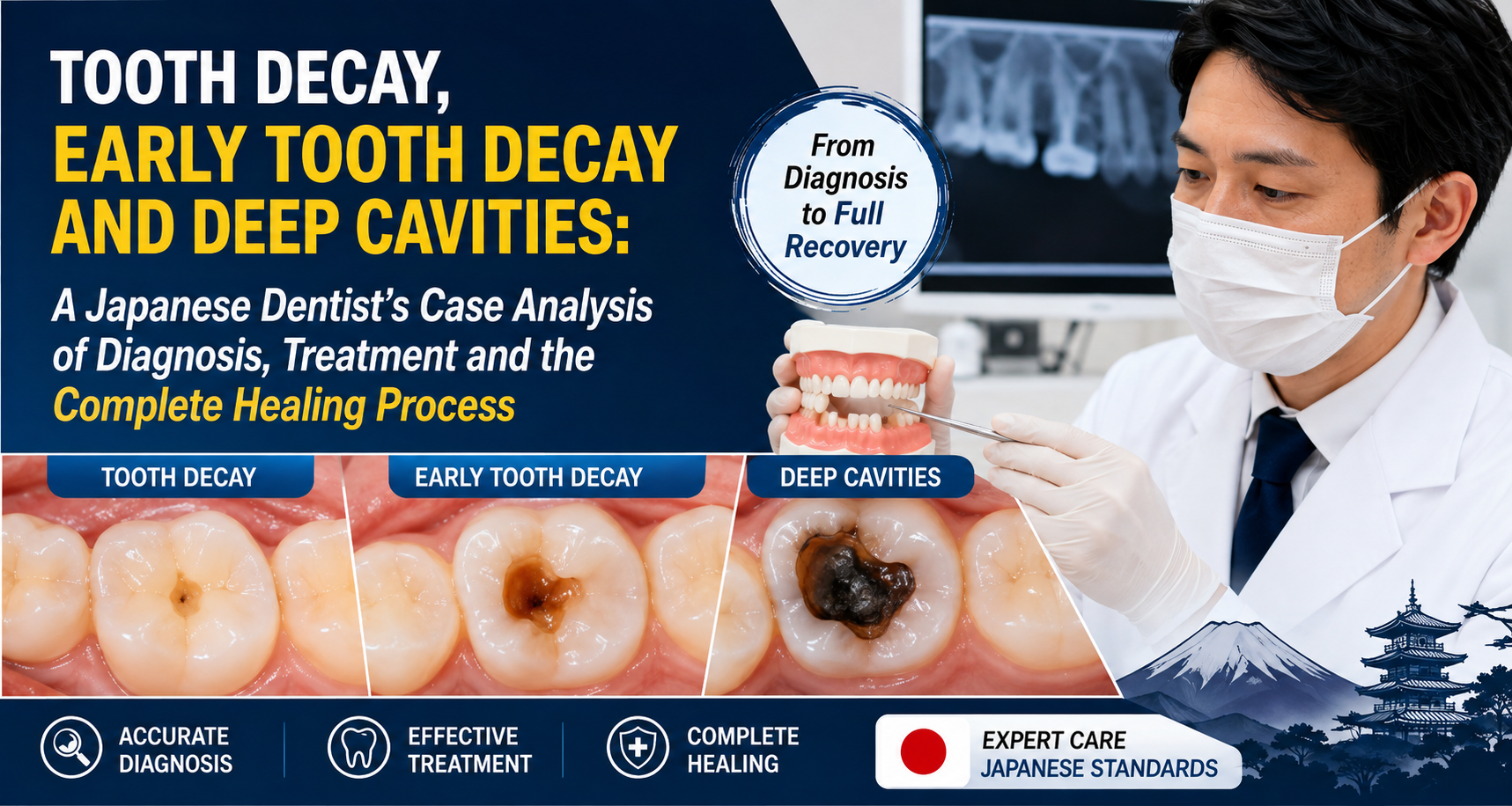
As a dentist practicing within the modern Japanese dental-care environment, I approach tooth decay as a disease process that can progress through different stages, rather than simply as a hole that needs to be drilled and filled.
This distinction is important.
A patient with an early, non-cavitated lesion may be managed very differently from a patient with a large cavity extending deeply toward the dental pulp.
Modern dentistry increasingly emphasizes early detection, prevention, remineralization when appropriate, preservation of healthy tooth structure, and conservative removal of diseased tissue when restoration becomes necessary. The American Dental Association (ADA) describes dental caries as a dynamic, multifactorial disease involving periods of demineralization and remineralization.
In this Dental Hub case analysis, I will review three important conditions:
-
Tooth Decay
-
Early Tooth Decay
-
Deep Cavities
I will also explain what I mean by the complete healing process—and an important point that patients sometimes misunderstand:
A tooth with early decay may be able to remineralize and become arrested, but a large cavitated hole generally does not simply grow back into normal tooth structure.
Treatment depends heavily on how advanced the lesion is.
Understanding Tooth Decay
Tooth decay is commonly called a cavity, but the terms are not exactly interchangeable.
Dental caries is the disease process.
A carious lesion is the resulting area of mineral loss and structural damage.
The ADA describes caries as a biofilm-mediated, sugar-driven, multifactorial and dynamic disease.
Several factors can contribute, including:
-
Dental plaque biofilm
-
Frequent sugar exposure
-
Poor oral hygiene
-
Reduced saliva
-
Certain medications
-
Dry mouth
-
Inadequate fluoride exposure
-
Deep grooves and pits
-
Previous history of cavities
-
Dietary patterns
-
Difficulty cleaning certain areas
This is why simply filling one cavity without addressing the patient's caries risk may not solve the underlying problem.
The Three Conditions We Are Reviewing
Condition 1: Tooth Decay
This is the broad category.
The tooth may have:
-
Demineralization
-
A visible lesion
-
A cavitated area
-
Dentin involvement
-
Deep caries
-
Possible pulpal involvement
The dentist determines the stage using clinical examination and, when appropriate, diagnostic imaging.
Condition 2: Early Tooth Decay
Early decay is particularly important because the tooth may still have substantial structural integrity.
A lesion may appear as:
-
A white or chalky area
-
A change in enamel translucency
-
Early discoloration
-
A non-cavitated lesion
At this stage, there may be no obvious hole.
This is where preventive and nonrestorative management can sometimes be extremely valuable.
Fluoride can promote remineralization of weakened enamel and help make the tooth surface more resistant to future acid attacks.
The ADA's clinical guidance includes nonrestorative approaches for certain noncavitated lesions, depending on the lesion's location, activity, and patient factors.
Condition 3: Deep Cavities
A deep cavity is substantially different.
The decay may extend through:
Enamel
↓
Dentin
↓
Deep dentin
↓
Near the pulp
↓
Possible pulpal inflammation or infection
At this stage, the dentist must balance two objectives:
-
Remove or control diseased tissue.
-
Preserve as much healthy tooth structure and pulp vitality as possible.
Modern restorative dentistry has moved away from the simplistic idea that every bit of softened or affected dentin must be aggressively removed until the dentist reaches hard tooth everywhere.
The ADA's 2023 restorative caries guideline supports conservative carious-tissue removal approaches, including selective caries removal for many moderate and advanced lesions in vital permanent teeth requiring restoration.
Case Analysis: A Patient in Japan With Three Stages of Decay
To understand the difference, consider a hypothetical 46-year-old patient visiting a dental clinic in Japan.
The patient reports:
"I don't have much pain, but my dentist said I have several cavities."
Clinical examination identifies three different types of lesions.
Tooth A
Small, non-cavitated enamel lesion.
Tooth B
Established cavity extending into dentin.
Tooth C
Large, deep cavity approaching the pulp.
Although all three may be described by the patient as "cavities," their management can be completely different.
Case 1: Early Tooth Decay
Clinical Finding
Tooth A has a localized chalky enamel area.
There is no obvious cavitation.
The patient has a history of frequent sweet snacks between meals.
The dentist assesses the patient's overall caries risk and determines that the lesion appears noncavitated.
What Happens Inside the Tooth?
At the microscopic level, acids produced by plaque bacteria can cause minerals to leave the enamel.
This is called demineralization.
But the process can move in the opposite direction.
Minerals can return to the enamel.
This is remineralization.
Fluoride supports this process and can help strengthen enamel against subsequent acid attacks.
Therefore, early decay is not necessarily an irreversible situation.
Early Decay: The Healing Process
When the lesion is appropriate for nonrestorative management, the goal is to shift the balance toward remineralization and disease control.
The process may include:
Step 1: Identify the lesion
The dentist determines whether the area is active, inactive, cavitated, or noncavitated.
Step 2: Assess caries risk
The dentist considers:
-
Diet
-
Plaque control
-
Fluoride exposure
-
Saliva
-
Previous cavities
-
Oral hygiene
-
Existing restorations
Step 3: Improve plaque control
The patient receives individualized oral-hygiene instruction.
Step 4: Control sugar exposure
Reducing frequent exposures to fermentable carbohydrates can reduce repeated acid attacks.
Step 5: Optimize fluoride exposure
Appropriate fluoride toothpaste and professionally recommended fluoride treatments may be used depending on age and caries risk.
The ADA identifies fluoride as an important tool for preventing and controlling dental caries.
Step 6: Monitor
The dentist reassesses the lesion.
The goal is not necessarily to make the previous white spot completely disappear.
The clinical objective may be to arrest the lesion and prevent further progression.
Can Early Tooth Decay Completely Heal?
This question requires careful wording.
Early, non-cavitated enamel lesions
These can potentially remineralize or become arrested when the disease process is controlled.
Established cavitated lesions
Once there is a true structural hole, the lost tooth structure generally does not regenerate biologically like skin or bone.
A restoration may be required to rebuild the missing structure.
This distinction is one of the most important concepts in modern minimally invasive dentistry.
Case 2: Established Cavity
Now consider Tooth B.
The patient has a visible cavity.
The lesion extends beyond enamel into dentin.
The tooth may be sensitive to:
-
Cold
-
Sweet foods
-
Certain acidic foods
The patient may not have spontaneous pain.
This is a classic example of why the absence of pain does not prove that the tooth is healthy.
What Happens When Decay Reaches Dentin?
Dentin is softer and structurally different from enamel.
Once the caries process reaches dentin, progression can potentially occur more rapidly.
The dentist evaluates:
-
Lesion depth
-
Cavitation
-
Tooth anatomy
-
Pulpal symptoms
-
Radiographic appearance
-
Restorability
-
Patient caries risk
Treatment may require a restoration.
Modern Treatment of Moderate Decay
The traditional concept was:
Drill until everything feels hard.
Modern evidence-based caries management can be more conservative.
For many vital permanent teeth requiring restoration, the ADA guideline recommends selective caries removal over more aggressive non-selective removal for moderate and advanced lesions.
The reasoning is important.
When decay is deep, aggressive removal may unnecessarily increase the risk of exposing the pulp.
The objective becomes:
Remove enough diseased tissue to create a cleanable, sealable restoration while preserving sound tooth structure and protecting the pulp whenever clinically appropriate.
The Healing Process After a Filling
Patients sometimes ask:
"Once I get the filling, is the tooth completely healed?"
I would explain it this way:
The cavity has been treated, but the tooth still requires protection.
After restoration:
-
The defective tooth structure is repaired.
-
The restoration seals the treated area.
-
The tooth remains under normal chewing forces.
-
The surrounding enamel and tooth surfaces still require protection.
-
The patient's caries risk still matters.
A filling does not make a person immune to future cavities.
Case 3: Deep Cavity
Now we reach the most clinically important case.
Tooth C has extensive decay extending deep toward the pulp.
The patient reports:
"It hurts when I drink something cold."
The pain disappears shortly after the cold stimulus is removed.
This information matters.
The dentist must determine whether the pulp is still capable of recovering or whether there is more advanced pulpal disease.
Deep Cavity Does Not Automatically Mean Root Canal
This is an important misconception.
A deep cavity does not automatically mean:
"The nerve is dead."
The dentist must evaluate:
-
Symptoms
-
Pulp testing
-
Percussion
-
Palpation
-
Radiographs
-
Caries depth
-
Remaining tooth structure
-
Restorability
A vital pulp may sometimes be preserved even when the cavity is deep.
This is one reason conservative caries removal is important.
The ADA specifically notes that conservative approaches may reduce adverse outcomes such as pulp exposure while preserving tooth structure.
Deep Caries: The Complete Treatment Process
Step 1: Diagnosis
The dentist determines the extent of decay.
Clinical examination and appropriate radiographs may be necessary.
Step 2: Pulp Assessment
The dentist evaluates whether the pulp appears:
-
Healthy
-
Reversibly inflamed
-
Irreversibly inflamed
-
Necrotic
The exact diagnosis depends on clinical findings.
Step 3: Determine Restorability
The dentist asks:
Can this tooth be predictably restored?
If sufficient healthy tooth structure remains, conservative restoration may be possible.
Step 4: Conservative Caries Removal
When clinically appropriate, the dentist may selectively remove carious tissue rather than aggressively excavating all tissue near the pulp.
The ADA's evidence-based guideline supports selective caries removal for many advanced lesions in vital permanent teeth requiring restoration.
Step 5: Protect the Pulp
Depending on the situation, the dentist may use an appropriate pulp-protective approach.
The specific material and technique depend on the clinical circumstances.
Step 6: Seal the Tooth
The remaining tooth structure is restored with an appropriate restorative material.
The goal is to create a durable seal and restore function.
Step 7: Monitor
The patient returns for appropriate follow-up.
The dentist monitors:
-
Symptoms
-
Restoration integrity
-
Tooth vitality when indicated
-
Radiographic changes when indicated
-
New caries risk
What If the Pulp Is Already Seriously Damaged?
Sometimes the decay has progressed too far.
The patient may have:
-
Spontaneous toothache
-
Severe or lingering thermal pain
-
Pain that wakes the patient
-
Pain when biting
-
Swelling
-
Tenderness
-
Evidence of infection
At that point, the treatment may involve endodontic therapy, extraction, or another appropriate approach depending on the diagnosis and restorability.
A deep cavity is therefore a situation where early professional evaluation is extremely important.
Case Review: Three Teeth, Three Different Strategies
| Condition | Typical Clinical Situation | Possible Approach |
|---|---|---|
| Early non-cavitated lesion | Enamel mineral loss without a hole | Risk control, fluoride/remineralization strategies, monitoring |
| Established cavity | Cavitation and dentin involvement | Restoration may be required |
| Deep cavity | Extensive decay approaching pulp | Conservative pulp-preserving approach when appropriate; endodontic treatment if pulp cannot recover |
These are general examples.
Actual treatment depends on the individual tooth and patient.
Why "Drill and Fill" Is No Longer the Whole Story
Modern caries management recognizes that drilling treats the lesion, but the disease process can continue elsewhere.
The ADA explicitly describes the older "drill and fill" paradigm as insufficient to address the full caries disease process, including the balance between demineralization and remineralization.
Imagine a patient who receives three fillings but continues to:
-
Sip sugary beverages throughout the day
-
Brush poorly
-
Have inadequate fluoride exposure
-
Have dry mouth
-
Develop new plaque accumulation
That patient can develop three more cavities.
The restorative treatment was successful.
The disease management was incomplete.
The Complete Healing Journey
A more accurate model is:
Stage 1 — Detection
The dentist identifies the lesion.
↓
Stage 2 — Diagnosis
The dentist determines its activity, depth, and clinical significance.
↓
Stage 3 — Risk Assessment
The dentist identifies why the patient is developing caries.
↓
Stage 4 — Disease Control
Diet, plaque control, fluoride exposure, saliva, and other risk factors are addressed.
↓
Stage 5 — Restorative Treatment When Necessary
Cavitated or structurally compromised lesions are restored.
↓
Stage 6 — Pulp Protection
Deep lesions are treated conservatively when appropriate.
↓
Stage 7 — Monitoring
The dentist checks whether the lesion and tooth remain stable.
↓
Stage 8 — Maintenance
The patient continues preventive care to reduce the risk of new disease.
This is what I would call the complete caries-management process.
Case Analysis: Six-Month Review
Return to our hypothetical patient.
At the initial visit:
Tooth A
Early noncavitated lesion.
Tooth B
Moderate cavity restored.
Tooth C
Deep cavity treated conservatively because the pulp remained clinically manageable.
Six months later, the patient returns.
Tooth A
The lesion appears stable.
No new cavitation is identified.
This is a successful outcome even if a slight visual discoloration remains.
Goal achieved: lesion arrested/stable.
Tooth B
The restoration is intact.
The patient has no symptoms.
Goal achieved: tooth restored and functioning.
Tooth C
The patient reports no spontaneous pain.
The tooth remains functional.
Clinical findings remain stable.
Goal achieved: tooth maintained without progression of symptoms.
However, continued monitoring remains important.
What Does "Complete Healing" Actually Mean?
For a cavity, "healing" should not always be interpreted as:
"The tooth becomes exactly like it was before the cavity."
For an early enamel lesion, remineralization and arrest can occur.
For a tooth requiring a filling, healing means controlling the disease and restoring the damaged structure.
For a deep cavity, healing may mean preserving a healthy or stable pulp and maintaining the tooth.
For a tooth requiring root canal treatment, the goal becomes controlling pulpal/periapical disease and restoring the tooth appropriately.
Therefore:
Healing is a clinical process, not simply the disappearance of a visible cavity.
Frequently Asked Questions
Can early tooth decay be reversed?
Early, noncavitated lesions can potentially remineralize or become arrested when the caries process is controlled. Fluoride is an important component of this process.
Can a deep cavity heal naturally?
A deep cavity with significant structural destruction generally cannot regenerate the lost tooth structure on its own. Professional treatment is usually required.
Does every cavity need a filling?
No.
Some early noncavitated lesions can be managed with nonrestorative approaches depending on their location, activity, and the patient's caries risk.
If my cavity does not hurt, do I still need treatment?
Possibly.
Caries can progress without significant pain, particularly during earlier stages.
Can a cavity reach the nerve without causing pain?
Yes.
Symptoms do not always correspond perfectly to the depth of a carious lesion.
Does a deep cavity always require a root canal?
No.
The dentist must evaluate the pulp and the overall condition of the tooth. Some deep lesions can be managed with conservative pulp-preserving treatment when the clinical situation permits.
What happens if the cavity reaches the pulp?
The pulp may become inflamed or infected. Depending on the diagnosis, treatment may include vital pulp therapy, root canal treatment, extraction, or another appropriate approach.
Can fluoride repair a large cavity?
Fluoride can support remineralization of early mineral loss, but it cannot rebuild a large structural hole in a tooth.
Can brushing remove a cavity?
Brushing can help control plaque and reduce future caries risk, but it cannot brush away an established structural cavity.
How long does a cavity take to heal after a filling?
A filling restores the damaged tooth, but the tooth does not become immune to future decay. Mild post-treatment sensitivity can occur, but persistent or worsening pain should be evaluated by the dentist.
Can early decay turn into a deep cavity?
Yes, if the caries process remains active and is not controlled.
How can I stop early decay from progressing?
Depending on your individual risk, your dentist may recommend improved plaque control, fluoride exposure, dietary changes, sealants for appropriate lesions, professional fluoride treatment, or other preventive strategies.
Is fluoride important for adults?
Yes. Fluoride can help remineralize weakened enamel and reduce susceptibility to acid attacks.
Can a dentist leave some decay under a filling?
In selected deep lesions, modern conservative approaches may intentionally avoid aggressive removal near the pulp. The ADA guideline supports selective caries removal in many moderate and advanced lesions requiring restoration.
Is leaving affected dentin dangerous?
The clinical objective is not simply to leave uncontrolled disease. The dentist uses an evidence-based caries-removal strategy and seals the tooth appropriately. The specific approach depends on lesion depth, pulp status, restorability, and other clinical factors.
Warning Signs That Require Prompt Dental Evaluation
Do not wait for a routine cleaning if you develop:
-
Persistent toothache
-
Severe sensitivity
-
Lingering pain after hot or cold
-
Pain when biting
-
Spontaneous pain
-
Facial swelling
-
Gum swelling near a tooth
-
Pus or drainage
-
Fever associated with dental symptoms
-
A broken tooth with deep decay
-
Difficulty opening your mouth
Facial swelling accompanied by difficulty breathing or swallowing requires urgent medical attention.
How Patients Can Support the Healing Process
The dentist can repair a tooth, but the patient plays a major role in preventing recurrence.
I recommend focusing on:
Brush twice daily with fluoride toothpaste
Fluoride helps strengthen enamel and supports remineralization.
Clean between teeth
Plaque can accumulate in areas a toothbrush cannot adequately reach.
Reduce frequent sugar exposure
The frequency of sugar and fermentable carbohydrate exposure matters because repeated acid attacks can favor demineralization.
Drink water regularly
This can support normal oral conditions, particularly when it replaces sugary beverages.
Address dry mouth
Reduced salivary flow can increase caries risk.
Attend dental examinations
Early lesions are much easier to manage than advanced cavities.
Follow your individualized fluoride recommendations
Patients at elevated caries risk may benefit from professionally applied or prescription-strength fluoride according to clinical judgment and established guidelines.
Japanese Dental Perspective on Minimally Invasive Dentistry
Japan has a strong tradition of preventive dentistry and long-term oral-health maintenance, and modern Japanese dental practice increasingly incorporates digital diagnostics, minimally invasive techniques, adhesive restorative materials, and preventive care.
The philosophy I would emphasize is:
Preserve the natural tooth whenever reasonably possible.
That does not mean avoiding necessary treatment.
It means avoiding unnecessary destruction of healthy tooth structure.
The modern evidence base supports this direction. The ADA's restorative guideline specifically emphasizes conservative approaches that can preserve healthy tooth structure and reduce risks associated with more aggressive caries removal.
Final Clinical Review
The three conditions in this case analysis illustrate why the phrase "I have a cavity" is not enough information for a dentist.
Early Tooth Decay
The tooth may still have an opportunity for remineralization or arrest.
Established Tooth Decay
The structural damage may require restoration.
Deep Cavity
The dentist must carefully evaluate the pulp and determine whether conservative treatment can preserve the tooth or whether more extensive treatment is necessary.
The most important lesson is that early diagnosis changes the treatment strategy.
A small noncavitated lesion may be managed conservatively.
A moderate cavity may require a restoration.
A deep cavity may require careful pulp-preserving treatment or, when the pulp is irreversibly damaged, endodontic treatment.
Modern dentistry is increasingly focused on controlling the disease while preserving as much healthy tooth structure as possible. The ADA's evidence-based recommendations support conservative caries-removal approaches for many moderate and advanced lesions in vital teeth requiring restoration.
The Complete Caries-Management Formula
Detect early
→ Assess risk
→ Control the disease
→ Remineralize when possible
→ Restore when necessary
→ Protect the pulp
→ Monitor healing
→ Prevent recurrence
That is the fundamental difference between simply fixing a cavity and providing comprehensive, modern caries management.
Dental Hub clinical note: This article is an educational case analysis, not a diagnosis. Actual treatment should be based on an examination, appropriate diagnostic testing, the patient's symptoms, caries risk, tooth anatomy, pulp status, and the dentist's clinical judgment.
Related Clinical Cases
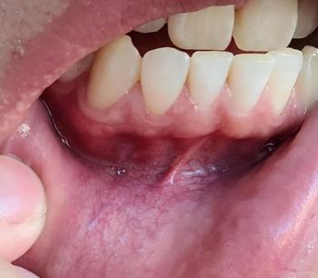
Patients with lower front gum inflammation may also experience tartar buildup, periodontal disease, gum abscesses, or tooth decay. Explore these related clinical case studies.


Other topics:
Other Dental Clinic