Dentures That Feel Natural, Look Beautiful.
Deep Dive: Clinical Analysis and Management of Periapical Lesions
Image:
Severity:
Teeth Problems:
Full Analysis and Diagnosis
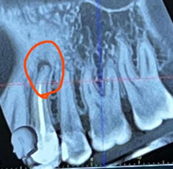
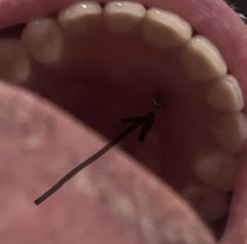
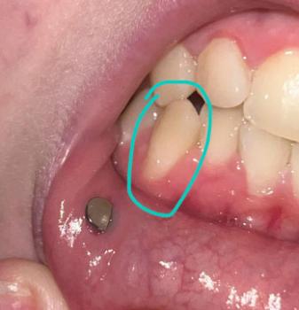
The provided image is a diagram or radiograph illustrating Periapical Lesions—pathological changes that occur around the apex (tip) of a tooth root, typically in response to a bacterial infection originating from the tooth's necrotic (dead) pulp.
-
Diagnosis: The general term for the condition shown is Chronic Apical Periodontitis (with a bone radiolucency), which can manifest histologically as a Periapical Granuloma, a Radicular Cyst, or a Periapical Abscess.
-
Cause: The lesion is a defensive mechanism (inflammation and bone destruction) caused by bacteria and their by-products leaking from the infected or necrotic pulp tissue within the root canal system. This usually results from deep dental decay (caries), trauma, or a failed/inadequate previous root canal treatment.
-
Scale All Teeth: The image shows a single tooth affected, but in a real clinical scenario, all teeth would need to be checked radiographically to ensure no other asymptomatic lesions are present.
Process to Execute (General Treatment Pathway)
The primary goal is to eliminate the source of infection within the tooth's root canal system to allow the surrounding bone to heal.
-
Phase 1: Diagnosis & Planning
-
Clinical Testing: Assess the tooth's vitality (it will typically be non-responsive to cold/electric pulp tests).
-
Imaging: Obtain a clear Periapical Radiograph (PA) or, ideally, a Cone-Beam Computed Tomography (CBCT) scan. The CBCT is superior for assessing the true size, nature (cyst vs. granuloma), and extent of the bone destruction, which guides the treatment.
-
-
Phase 2: Definitive Treatment (Non-Surgical)
-
Root Canal Treatment (RCT): The standard of care. This involves:
-
Accessing the pulp chamber and root canals.
-
Removing all infected/necrotic tissue.
-
Thorough cleaning, shaping, and disinfection of the entire canal system using specialized instruments and irrigating solutions (e.g., Sodium Hypochlorite).
-
Placing an intracanal medication (e.g., Calcium Hydroxide) for a period, often weeks or months, to further sterilize the canals, especially in larger lesions.
-
Obturation: Sealing the root canals with filling material (e.g., Gutta-percha and sealer) to prevent re-infection.
-
-
-
Phase 3: Restoration
-
The tooth must be immediately restored with a proper seal (e.g., filling) and often a Dental Crown to protect the weakened tooth structure from fracture.
-
-
Phase 4: Follow-up & Monitoring
-
The patient must be recalled for clinical and radiographic follow-up at 6, 12, and 24 months to monitor the bone healing.
-
Time Frame to Heal
Healing is a bone regeneration process, not a 14-day event.
-
Active Treatment: The root canal procedure itself may take 1 to 3 visits over a few days to weeks.
-
Healing Time Frame: The bone defect (the periapical lesion) takes a long time to fill in and regenerate.
-
Initial Signs of Healing: Often visible on X-ray at 6 to 12 months.
-
Complete Healing: Can take 1 to 4 years or more, depending on the original size of the lesion, whether it was a granuloma or a cyst, and the patient's general health.
-
If it Takes 14 Days
-
No Healing is Expected: In 14 days, the patient should expect relief from acute pain (if an abscess was present) and the completion of the initial non-surgical Root Canal Treatment (the cleansing and sealing phase).
-
Issue that Will Scale Up: If the patient stops treatment after 14 days without completing the full obturation and final restoration (crown):
-
Coronal Leakage: The temporary filling will fail, allowing bacteria from the mouth to seep back into the freshly cleaned root canals.
-
Re-infection: The entire root canal system will become contaminated again.
-
Treatment Failure: The lesion will persist or grow, leading to the eventual need for Endodontic Retreatment, Surgery (Apicoectomy), or Tooth Extraction.
-
Comments
This is a common but serious dental condition. The successful outcome hinges on meticulous root canal disinfection. The healing of the bone is entirely dependent on the complete elimination of bacteria from the canal system. If the tooth remains symptomatic after the non-surgical RCT, or if the lesion shows no radiographic reduction after 1-2 years, surgical intervention (apicoectomy or tooth extraction) would be the next step.
Visit Nearest Location Area
To find a qualified endodontist or general dentist specializing in root canal therapy who can properly treat this condition, please use the provided directory link:
Directory Listing: https://cebudentalimplants.com/map-dental-clinic
Teeth Case
 |
 |
 |
 |
 |
 |


