Your Smile, Our Passion.

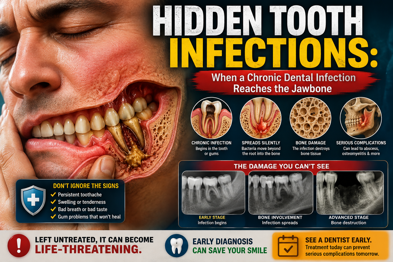
Hidden Tooth Infections: When a Chronic Dental Infection Reaches the Jawbone
image hero:
Dental Hub Clinical Analysis
Expert perspective: USA dental clinical voice
Topic: Chronic Tooth Infection and Osteomyelitis of the Jaw
Format: Case Study + Clinical Analysis + Diagnostic Reasoning
Language: English
Introduction: The Infection You Cannot Always See
A tooth infection does not always announce itself with severe pain.
Some chronic dental infections develop quietly. The patient may experience only occasional sensitivity, bad taste, localized gum swelling, intermittent discomfort, or no obvious symptoms at all. Meanwhile, bacteria associated with an infected tooth can persist around the root and, in certain circumstances, extend into surrounding tissues and jawbone.
One of the more serious complications is osteomyelitis, an infection and inflammatory process involving bone.
In the jaws, osteomyelitis can develop from an untreated or inadequately controlled odontogenic infection. Published case reports describe chronic jaw osteomyelitis associated with dental infections, with findings that can include swelling, drainage, fistula formation, pain, sensory changes, bone destruction, sclerosis, and areas of devitalized bone.
The important clinical lesson is simple:
A tooth can look relatively normal from the outside while the pathology is occurring underneath the gum and inside the bone.
Case Study: The “Toothache That Went Away”
Patient Profile
Consider this representative clinical scenario:
A 52-year-old patient presents to a dental office with a history of intermittent discomfort involving a lower molar.
The patient explains:
“It used to hurt, but the pain disappeared. I thought the infection was gone.”
There is no dramatic facial swelling.
There is no high fever.
The patient is eating normally.
However, clinical examination reveals:
-
A heavily restored lower molar
-
Localized tenderness
-
Mild swelling around the tooth
-
A draining point on the gum
-
Slight tooth mobility
-
A history of recurrent episodes of discomfort
The patient's previous dental records show that the tooth had experienced extensive decay and previous dental treatment.
At this point, the clinician should not assume that the absence of severe pain means the infection has resolved.
Step 1: Why the Pain Can Disappear
This is one of the most misunderstood aspects of chronic tooth infection.
When the pulp inside a tooth becomes severely inflamed or dies, the nerve may lose its ability to generate the intense pain the patient previously experienced.
That does not necessarily mean the infection has disappeared.
Instead, bacteria and inflammatory products may continue to move through the root canal system toward the tissues surrounding the root.
The clinical picture can therefore change from:
severe tooth pain → reduced pain → intermittent symptoms → chronic infection
This is why a patient saying, “It stopped hurting,” should not automatically be interpreted as “the tooth healed.”
Step 2: The Hidden Infection Beneath the Tooth
The next question is:
Where is the infection actually located?
A dental infection can involve several anatomical compartments:
-
The tooth's pulp
-
The root canal system
-
The periodontal ligament
-
The tissues around the root apex
-
The cortical bone
-
The cancellous or medullary bone
-
Adjacent facial spaces
When infection extends beyond the tooth and surrounding periodontal tissues, the clinical problem becomes considerably more complex.
Jaw osteomyelitis is an uncommon but potentially serious condition. It can occur through contiguous spread from odontogenic infection, although jawbone infection can also have other causes such as trauma, surgery, fractures, radiation, or other disease processes.
Step 3: The Imaging Changes the Diagnosis
The patient receives dental radiographs.
The clinician identifies an abnormal area around the affected tooth.
Instead of seeing only a straightforward localized periapical lesion, the imaging raises additional questions:
-
Is there destruction of the surrounding bone?
-
Is there sclerosis?
-
Is there a sequestrum?
-
Is the lesion confined to the tooth apex?
-
Has the process extended beyond the immediate periapical region?
-
Could another pathology mimic an infection?
A CBCT scan may provide additional three-dimensional information when conventional imaging does not adequately define the extent of disease.
In published cases of chronic jaw osteomyelitis, imaging modalities including conventional radiography, CT, MRI, and other investigations have been used alongside clinical and pathological evaluation.
Step 4: The Critical Clinical Distinction
At this stage, the dentist must distinguish between several possibilities.
Localized dental infection
The infection may remain primarily associated with the tooth and surrounding periapical tissues.
Chronic odontogenic infection
A persistent source may remain inside the tooth or surrounding periodontal structures.
Osteomyelitis
The infection and inflammatory process has extended into the jawbone.
Noninfectious inflammatory bone disease
Not every case of jaw inflammation is caused by bacteria. Primary chronic osteomyelitis and other inflammatory conditions can produce overlapping symptoms and imaging findings.
Other pathology
Tumors, cystic lesions, medication-related osteonecrosis, traumatic lesions, and other disorders may sometimes mimic infection.
This is why a diagnosis should never be based on a single symptom or one radiographic image.
Step 5: Clinical Red Flags for a Deeper Problem
A dentist should take persistent or recurrent symptoms seriously when they include:
-
Recurrent swelling
-
Persistent drainage
-
A gum fistula
-
Pus or foul-tasting drainage
-
Persistent localized pain
-
Facial asymmetry
-
Unexplained tooth mobility
-
Difficulty opening the mouth
-
Altered sensation or numbness
-
Nonhealing areas following extraction
-
Recurrent infection in the same region
-
Abnormal bone destruction or sclerosis on imaging
Published chronic mandibular osteomyelitis cases have reported combinations of pain, swelling, purulent discharge, fistulas, sensory disturbances, trismus, and pathological fracture.
Importantly, not every patient will have all of these findings.
Step 6: Why Antibiotics Alone May Not Solve the Problem
This is a critical point in modern dental infection management.
An antibiotic can help control bacterial infection in appropriate circumstances, but it does not automatically eliminate the underlying dental source.
For common pulpal and periapical dental conditions, the American Dental Association emphasizes definitive dental treatment rather than routine antibiotic use. Depending on the diagnosis, this can include procedures such as root canal treatment or drainage; antibiotics become particularly important when systemic involvement is present.
With suspected jaw osteomyelitis, management becomes more complex and may require coordinated evaluation by dental specialists, oral and maxillofacial surgeons, infectious-disease specialists, radiologists, pathologists, and microbiologists depending on the case.
Published cases demonstrate that treatment can involve antimicrobial therapy and, when indicated, surgical removal of infected or nonviable tissue.
The principle is:
Control the infection and identify the source.
Step 7: When Dead Bone Becomes Part of the Problem
Chronic osteomyelitis can produce areas of devitalized bone known as sequestra.
These pieces of nonviable bone can become a persistent focus for inflammation and recurrent infection.
A published case of chronic suppurative mandibular osteomyelitis described CBCT evidence of a bone sequestrum followed by surgical removal of the affected bone and debridement.
This demonstrates why a persistent bone infection cannot always be approached as though it were simply another toothache.
The clinician must determine:
Where is the infection?
What is maintaining it?
Is there nonviable tissue?
Is the original tooth still the source?
Step 8: The Role of Microbiology and Biopsy
In complicated cases, identifying the responsible organism may be difficult.
A superficial swab does not necessarily provide the same information as properly collected deep tissue or bone specimens.
One published case of chronic bacterial osteomyelitis of the jaw required repeated microbiological sampling before advanced laboratory analysis identified bacterial organisms. The final diagnosis depended on integrating clinical findings, imaging, pathology, and microbiology.
This illustrates an important principle:
The diagnosis of complex jaw infection is multidisciplinary.
Clinical examination alone may not tell the entire story.
Step 9: The Differential Diagnosis Matters
An experienced clinician should avoid labeling every abnormal jaw lesion as “chronic infection.”
The differential diagnosis may include:
-
Chronic odontogenic infection
-
Acute or chronic osteomyelitis
-
Primary chronic osteomyelitis
-
Osteonecrosis
-
Medication-related osteonecrosis of the jaw
-
Odontogenic cysts
-
Odontogenic tumors
-
Malignancy
-
Chronic sinus disease involving the maxilla
-
Traumatic bone injury
-
Other inflammatory bone disorders
Medication-related osteonecrosis is particularly important to distinguish from infection because it has specific diagnostic criteria and associations with medications such as antiresorptive and antiangiogenic therapies.
Case Study: Clinical Reasoning
Returning to our representative patient, the dentist now has three major pieces of information:
Clinical examination
There is recurrent localized swelling and drainage.
Dental history
The affected molar has a significant history of decay and previous treatment.
Imaging
The bone surrounding the tooth demonstrates an abnormal pattern that extends beyond a simple localized lesion.
The clinician now considers:
Chronic odontogenic infection with possible extension into the jawbone.
At this point, referral to an appropriate dental specialist or oral and maxillofacial surgeon may be warranted for further evaluation.
Additional imaging, laboratory testing, microbiological sampling, biopsy, or surgical assessment may be considered depending on the clinical presentation.
The Biggest Diagnostic Trap
The most dangerous assumption is:
“If the tooth doesn't hurt anymore, the infection must be gone.”
That conclusion is not clinically reliable.
Pain depends on many factors, including the status of the dental pulp, pressure within tissues, drainage pathways, and nerve function.
A chronic infection may therefore produce relatively mild symptoms while significant pathology remains.
The opposite is also true:
Severe pain does not automatically mean osteomyelitis.
Diagnosis requires clinical examination and appropriate investigation.
When a Dental Infection Becomes an Emergency
A patient with a suspected dental infection should seek urgent professional evaluation if symptoms are rapidly worsening.
Particularly concerning features include:
-
Rapidly increasing facial swelling
-
Swelling extending toward the neck
-
Difficulty breathing
-
Difficulty swallowing
-
Significant difficulty opening the mouth
-
Fever or systemic illness
-
Rapid deterioration
-
Confusion or profound weakness
Systemic involvement changes the clinical urgency. The ADA's dental infection guidance specifically recognizes fever and malaise as signs that can warrant antibiotic therapy in appropriate dental infections, alongside definitive dental care.
Airway compromise is an emergency requiring immediate medical attention.
Clinical Analysis: What This Case Teaches
1. Pain is not a reliable infection detector
A chronic infection can exist with surprisingly little pain.
2. A draining fistula is not necessarily a sign of healing
Drainage can reduce pressure and pain while the underlying source remains.
3. Imaging provides information that the mouth cannot reveal
The external appearance of the gum may underestimate the extent of underlying bone involvement.
4. Osteomyelitis is a diagnosis that requires careful investigation
Clinical examination, imaging, microbiology, and pathology may all contribute.
5. Antibiotics are not a substitute for source control
The underlying dental source must be addressed when appropriate.
6. Not every bone lesion is bacterial osteomyelitis
Noninfectious inflammatory diseases and other jaw pathologies can mimic infection.
7. Persistent disease deserves escalation
When symptoms repeatedly return despite initial treatment, the diagnosis and treatment strategy should be reassessed.
Final Dental Hub Perspective
Hidden dental infections are important because they challenge the patient's perception of what “infection” looks like.
A patient may have:
little pain + little swelling + apparently normal chewing
while an underlying dental infection remains active.
In more advanced cases, infection can extend into the jawbone and produce osteomyelitis, with potentially serious consequences.
The appropriate response is not panic.
It is accurate diagnosis, identification of the infection source, appropriate imaging, and timely specialist management when indicated.
If a tooth repeatedly becomes painful, swollen, drains through the gum, fails to heal after treatment, or produces unexplained changes on dental imaging, the problem deserves a comprehensive evaluation rather than repeated temporary treatment.
The absence of pain does not prove the absence of disease.
And when bone involvement is suspected, the question is no longer simply:
“Which tooth hurts?”
The more important question becomes:
“What is happening beneath the tooth—and has the infection reached the bone?”
Medical Disclaimer
This Dental Hub case study is for educational purposes and represents a clinical teaching scenario rather than a diagnosis of an actual patient. Osteomyelitis of the jaw has multiple causes and can resemble other dental, inflammatory, or bone disorders. Diagnosis and treatment require evaluation by a qualified dental or medical professional. Do not start, stop, or change antibiotics without professional guidance.
Related Clinical Cases
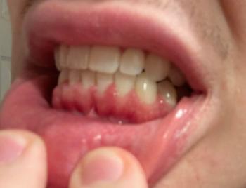
Patients with lower front gum inflammation may also experience tartar buildup, periodontal disease, gum abscesses, or tooth decay. Explore these related clinical case studies.
Other topics:
Other Dental Clinic