Your Smile, Our Passion.

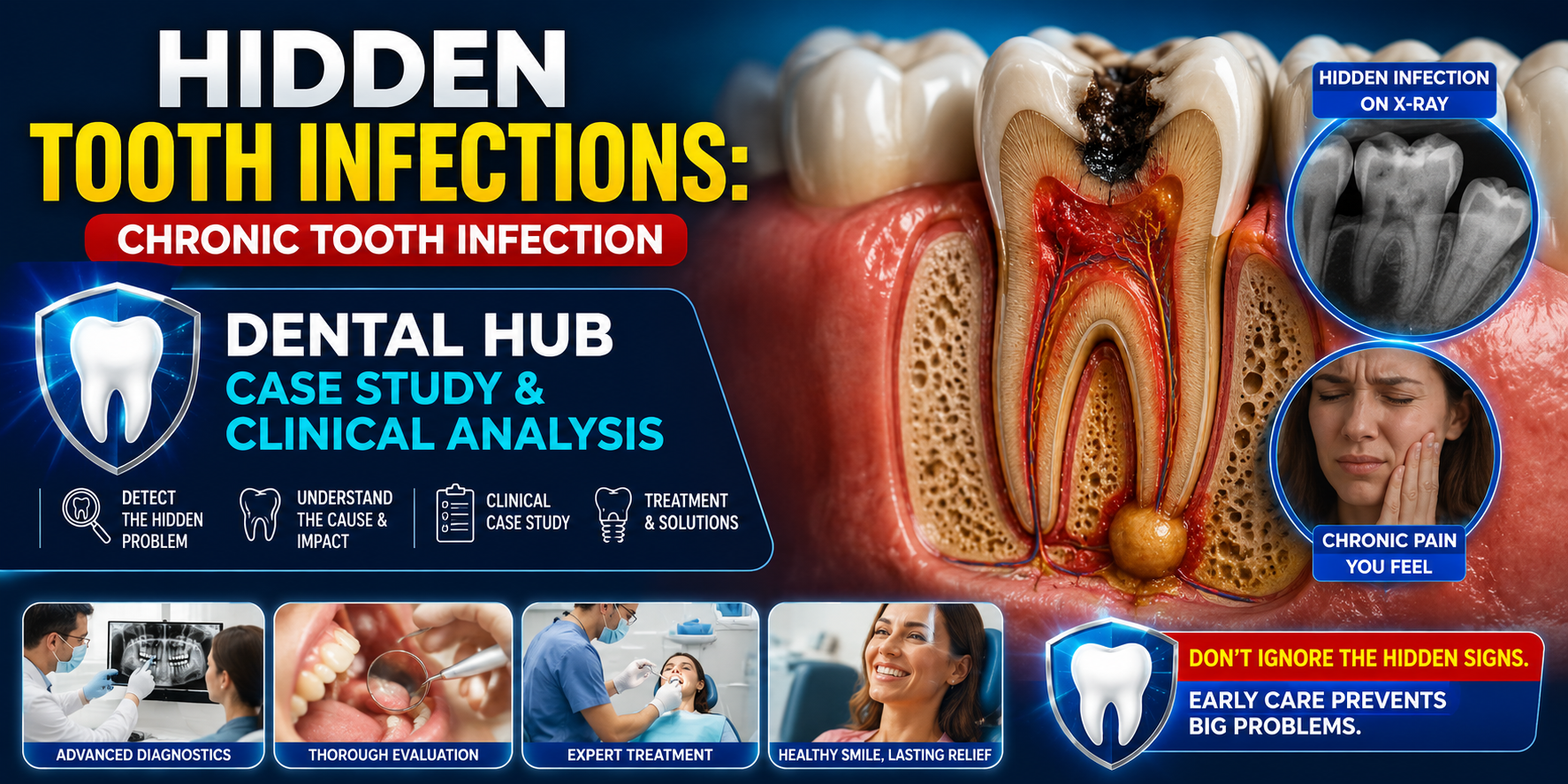
Hidden Tooth Infections: Chronic Tooth Infection Dental Hub Case Study & Clinical Analysis
image hero:
Country:
A tooth can look relatively normal on the outside while a chronic infection continues to develop deep inside the tooth or around its roots. These hidden tooth infections can be difficult to recognize because pain may be mild, intermittent, or completely absent.
From a U.S. dental clinical perspective, the important lesson is simple: absence of severe pain does not necessarily mean absence of infection.
This case-study analysis examines how a chronic tooth infection can develop, why symptoms may disappear, how dentists investigate the problem, and how treatment decisions are made.
Case Study: The Patient Who Had Almost No Pain
A 48-year-old patient presented for a routine dental examination. The patient reported occasional sensitivity when chewing but denied significant toothache, facial swelling, or fever.
The tooth looked relatively intact clinically.
There was no obvious large cavity visible during the initial examination.
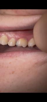
However, the dentist noticed a subtle change in the gum tissue near the tooth. The area was slightly tender to pressure, and the patient reported that a small pimple-like bump had occasionally appeared and disappeared over several months.
This raised suspicion of a possible chronic infection.
Initial Clinical Findings
The dentist evaluated:
-
Tooth decay and previous restorations
-
Gum condition around the tooth
-
Tenderness to biting or percussion
-
Tooth mobility
-
Response to cold or other vitality testing
-
Periodontal probing depths
-
Presence of drainage or a sinus tract
-
Adjacent teeth for comparison
The clinical findings alone did not completely explain the problem.
That is where dental imaging became important.
The Hidden Problem: What the X-Ray Revealed
A periapical dental radiograph showed a subtle dark area around the root tip.
This type of radiolucency can indicate inflammatory changes associated with infection originating from the tooth's pulp, although radiographic findings must always be interpreted together with the clinical examination.
The patient was surprised.
There had been no severe toothache.
This is one of the most important concepts in chronic dental infection:
The intensity of pain does not reliably measure the seriousness or extent of a dental infection.
When the pulp becomes necrotic, the nerve tissue inside the tooth may no longer respond normally to stimulation. Pressure from an infection can also periodically drain through the gum, reducing symptoms.
The result can be a quiet chronic infection.
How a Chronic Tooth Infection Develops
The process often begins with damage to the tooth.
Common pathways include:
1. Deep Dental Decay
Bacteria progressively penetrate through enamel and dentin.
Eventually, the infection can reach the dental pulp.
The pulp contains nerves and blood vessels. Once significant inflammation or bacterial contamination occurs, the pulp may become irreversibly inflamed and eventually necrotic.
2. Cracked or Fractured Teeth
A crack can provide a pathway for bacteria.
Some cracks are difficult to see clinically, particularly when they occur beneath an existing restoration.
3. Previous Dental Treatment
A tooth that previously received a filling, crown, or root canal may develop problems later.
For root-canal-treated teeth, persistent or recurrent disease can occur for several reasons, including residual infection, missed anatomy, inadequate cleaning or sealing, or new leakage.
4. Trauma
A tooth that suffered trauma years earlier can sometimes develop pulp necrosis long after the original injury.
The patient may not associate the current dental problem with an old accident.
Why the Infection Can Become "Hidden"
The body's inflammatory response does not always produce dramatic symptoms.
A chronic infection may establish a relatively stable drainage pathway through the bone and gum.
This can produce:
-
A small gum bump
-
Occasional drainage
-
Bad taste
-
Mild tenderness
-
Intermittent discomfort
-
Discomfort when biting
-
Darkening of a tooth
-
No symptoms at all
The infection may therefore be discovered incidentally during an examination or dental imaging.
Clinical Analysis: What the Dentist Must Determine
A responsible diagnosis should not be based on an X-ray alone.
The dentist must determine several things.
Is the Tooth Vital?
Pulp vitality or sensibility testing helps determine whether the nerve tissue is responding.
A nonresponsive tooth may suggest pulp necrosis, but test results must be interpreted carefully because no single test is perfect.
Is There Apical Inflammation?
The dentist evaluates the tissues around the root.
Tenderness to biting or percussion can provide additional evidence of inflammation involving the periodontal ligament.
Is There a Periodontal Problem?
Deep periodontal pockets can sometimes mimic or contribute to endodontic disease.
The dentist therefore performs periodontal probing around the tooth.
Is There a Crack?
A vertical root fracture or significant tooth crack can fundamentally change the prognosis and treatment strategy.
This is one reason a tooth should not automatically be treated with root canal therapy simply because an X-ray shows a dark area.
Advanced Imaging: When CBCT Becomes Important
In selected cases, a dentist may recommend cone-beam computed tomography (CBCT).
CBCT can provide three-dimensional information that conventional two-dimensional radiographs cannot fully demonstrate.
It may help evaluate:
-
Root anatomy
-
Periapical bone changes
-
Additional canals
-
Complex anatomy
-
Relationship to nearby structures
-
Suspected root fractures
-
Persistent disease after previous treatment
CBCT is not automatically necessary for every dental infection. The dentist weighs the diagnostic benefit against radiation exposure and uses it when the additional information is clinically justified.
Case Study Turning Point
Returning to our patient, the dentist discovered that the affected tooth had previously undergone extensive restoration.
The tooth responded poorly to vitality testing compared with neighboring teeth.
The radiograph showed a periapical lesion.
Further evaluation suggested that the tooth had a chronic endodontic infection.
The absence of severe pain had created a false sense of security.
The treatment discussion then focused on whether the tooth could be predictably saved.
Treatment Decision: Save the Tooth or Remove It?
The treatment depends on the condition of the tooth, the source of infection, periodontal support, restorability, and the patient's overall circumstances.
Root Canal Treatment
If the tooth is restorable and the infection originates from the pulp, root canal treatment may be appropriate.
The objective is to:
-
Remove infected or necrotic pulp tissue.
-
Clean and shape the root canal system.
-
Disinfect the canals.
-
Seal the canal system.
-
Restore the tooth to prevent reinfection.
Root Canal Retreatment
If a previously treated tooth develops persistent or recurrent disease, retreatment may be considered when the tooth is restorable and the cause of failure can potentially be corrected.
Endodontic Surgery
In selected cases, an endodontic surgical procedure such as an apicoectomy may be considered when conventional retreatment is not appropriate or has not resolved the disease.
Extraction
Extraction may become necessary when a tooth cannot be predictably restored.
Examples can include certain severe fractures, inadequate remaining tooth structure, or an unfavorable periodontal or structural prognosis.
Extraction is not automatically the best treatment simply because infection is present.
What About Antibiotics?
This is an important clinical distinction.
Antibiotics do not replace definitive dental treatment for most localized tooth infections.
The source of infection generally needs to be addressed through appropriate dental treatment such as drainage, root canal treatment, or extraction.
Antibiotics may be indicated when there are systemic signs or spreading infection, depending on the clinical situation.
A dentist or physician should determine whether antibiotics are appropriate.
Red Flags: When a "Hidden" Infection Becomes an Emergency
A chronic infection can sometimes become acutely worse.
Seek urgent professional evaluation when symptoms include:
-
Rapidly increasing facial swelling
-
Swelling around the eye
-
Difficulty swallowing
-
Difficulty breathing
-
Significant difficulty opening the mouth
-
Fever or systemic illness
-
Rapid progression of swelling
-
Severe uncontrolled pain
-
Significant weakness or feeling seriously ill
Difficulty breathing or swallowing, or rapidly progressive swelling involving the face or neck, can represent a medical emergency.
The Dental Hub Clinical Takeaway
This case demonstrates why routine dental examinations matter.
A chronic tooth infection may not behave like the stereotypical dental abscess.
There may be:
No dramatic pain.
No obvious cavity.
No major swelling.
Yet the tooth may still have significant disease.
The combination of clinical examination, pulp testing, periodontal evaluation, and appropriate dental imaging allows the dentist to determine what is actually happening.
The goal is not simply to find a dark area on an X-ray.
The goal is to identify the source of disease, determine whether the tooth is restorable, and choose treatment that offers the patient the best predictable outcome.
Final Clinical Perspective
Hidden tooth infections are not necessarily "silent killers," but they should not be ignored.
A chronic dental infection can persist with surprisingly few symptoms. A small recurring gum bump, unexplained bad taste, tenderness when chewing, or a tooth that changes color can justify a professional dental evaluation.
The most important lesson from this case study is:
Don't use pain alone as your measure of dental health.
A tooth that feels fine may still require investigation.
For patients in the United States, the appropriate next step is a comprehensive examination by a licensed dentist, with referral to an endodontist when the case involves complex root-canal disease or diagnosis.
Dental Hub Clinical Series:
Hidden Tooth Infections — Chronic Tooth Infection: Case Study, Diagnostic Reasoning, Imaging, Treatment Options, and Emergency Warning Signs.
Related Clinical Cases
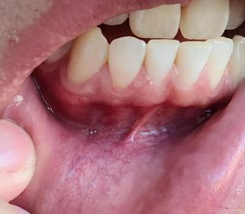
Patients with lower front gum inflammation may also experience tartar buildup, periodontal disease, gum abscesses, or tooth decay. Explore these related clinical case studies.



Other topics:
Other Dental Clinic