Dentures That Feel Natural, Look Beautiful.

Dental Case Analysis: Heavy Lower-Tooth Calculus, Possible Gingival Disease, and a Floor-of-Mouth Lesion
Image:
Severity:
Teeth Problems:
Additional Clinical Insights: Heavy Tartar on Lower Teeth, Gum Disease Risk, and a Lesion Beneath the Tongue
1. Why Does Tartar Accumulate Behind the Lower Front Teeth?
The inner surfaces of the lower front teeth are particularly susceptible to calculus accumulation because the openings of the submandibular and sublingual salivary ducts are located nearby. Saliva contains minerals that can help harden dental plaque into calculus over time.
Once plaque mineralizes, ordinary brushing cannot reliably remove the hardened deposits. Professional dental scaling is needed to remove them.
Crowded teeth, inadequate interdental cleaning, irregular dental visits, and individual differences in plaque accumulation can contribute to the problem.
Clinical point: Heavy calculus is a reason to investigate gum health, but it does not automatically mean that the patient has advanced periodontitis or bone loss.
2. Can Heavy Calculus Cause Gum Disease?
Yes. Calculus creates a rough surface that retains plaque, making effective cleaning more difficult. The bacteria in dental plaque can trigger inflammation of the surrounding gingiva.
Two conditions must be distinguished:
-
Gingivitis: Gum inflammation characterized by findings such as redness, swelling, and bleeding. It does not involve the attachment and supporting-bone destruction that defines periodontitis.
-
Periodontitis: A disease involving destruction of the tissues supporting the teeth. It can cause periodontal pocket formation, attachment loss, and bone loss.
A dentist distinguishes these conditions through periodontal probing, assessment of clinical attachment levels, and dental radiographs when indicated.
3. Why Is the Floor-of-Mouth Lesion Important?
A raised or discolored area beneath the tongue should not automatically be attributed to tartar or gum disease.
The floor of the mouth contains important anatomical structures, including salivary glands, salivary ducts, blood vessels, and soft tissues. A visible prominence may represent normal anatomy, local irritation, a salivary-duct problem, a cyst, or another oral lesion.
A photograph alone cannot reliably distinguish these possibilities.
During an examination, the dentist should assess the lesion's location, size, color, consistency, tenderness, duration, and whether it changes during meals. Salivary-duct obstruction may cause swelling or discomfort that becomes more noticeable when eating.
An unexplained lump, persistent ulcer, red or white patch, or lesion that is enlarging should receive prompt dental assessment. A mouth ulcer lasting more than three weeks, or a persistent lump or patch, warrants professional evaluation. These findings do not necessarily indicate cancer, but they should not be ignored.
4. What Is the Appropriate Treatment Plan?
Treatment should follow the confirmed clinical diagnosis rather than the photograph alone.
Step 1: Comprehensive dental examination
The dentist evaluates the teeth, gums, tongue, and floor of the mouth. Periodontal measurements help establish whether the problem is limited to gingivitis or includes deeper periodontal destruction.
Step 2: Professional calculus removal
Scaling removes hardened deposits from the tooth surfaces. If periodontal pockets and subgingival deposits are present, scaling and root planing may be indicated.
Step 3: Assessment of the floor-of-mouth lesion
The dentist determines whether observation, treatment of an identifiable cause, further investigation, or specialist referral is appropriate. Persistent or suspicious lesions may require additional diagnostic procedures, including biopsy when clinically indicated.
Step 4: Home plaque control
Brush twice daily with fluoride toothpaste, clean between the teeth daily, and follow the dentist's personalized oral hygiene instructions.
Step 5: Follow-up
The dentist reassesses gum inflammation and periodontal findings after treatment. Patients with periodontitis may require ongoing periodontal maintenance rather than a single cleaning.
5. When Is This an Emergency?
Seek emergency medical care if swelling beneath the tongue or in the floor of the mouth is rapidly increasing, particularly when accompanied by difficulty breathing, difficulty swallowing saliva, or elevation of the tongue.
Fever, spreading facial or neck swelling, or rapidly worsening pain also warrants urgent assessment.
Do not attempt to drain, puncture, or scrape a lesion yourself, and do not take antibiotics without appropriate medical or dental evaluation.
Frequently Asked Questions
1. Can tartar behind the lower front teeth cause bleeding gums?
Yes. Calculus can retain plaque and make cleaning difficult, contributing to gingival inflammation and bleeding. Persistent bleeding should be evaluated because periodontitis or other conditions may also be present.
2. Can brushing remove heavy dental calculus?
No. Brushing and interdental cleaning help remove plaque and reduce new accumulation, but hardened calculus generally requires professional removal with appropriate dental instruments.
3. Does heavy tartar always mean periodontitis?
No. Calculus can occur with gingivitis or periodontitis. A dentist must assess periodontal pocket depths, attachment loss, and supporting-bone levels before diagnosing periodontitis.
4. Can gum disease cause a lump beneath the tongue?
Not necessarily. A floor-of-mouth lump may involve salivary tissue, a duct, local irritation, a cyst, or another condition. It should be examined independently rather than assumed to be caused by gum disease.
5. How long do gums take to improve after scaling?
Gingival inflammation may begin improving within several days, with further improvement over the following one to two weeks when plaque control is effective. The timeline varies according to disease severity and the patient's response. Established periodontitis requires longer-term management.
6. Is full-mouth scaling necessary if only the lower front teeth have heavy tartar?
Not automatically. The dentist should examine the entire mouth and determine which areas need treatment. Full-mouth scaling may be appropriate when deposits or periodontal inflammation are more widespread.
7. Will scaling make teeth loose?
Scaling does not ordinarily cause healthy teeth to become loose. However, removing heavy calculus can reveal mobility that was previously masked by deposits. If a tooth is mobile, the dentist should investigate possible attachment loss, bone loss, or other causes.
8. Can I use a metal tool to remove tartar at home?
No. Attempting to scrape calculus yourself can injure the gums, damage tooth surfaces, or cause bleeding. Professional instruments and appropriate techniques should be used by a dental professional.
9. When should a lump beneath the tongue be checked?
Arrange a dental assessment promptly for an unexplained lump, particularly if it is enlarging, firm, painful, ulcerated, bleeding, or associated with difficulty swallowing. Do not wait three weeks to seek an appointment for a concerning lesion. A lesion that persists for more than three weeks requires evaluation even if it is not painful.
10. Can a salivary-duct problem cause swelling beneath the tongue?
Yes. A blocked or inflamed salivary duct can cause localized swelling and sometimes pain, particularly around meals. A dentist or appropriate medical specialist can assess the area and determine whether further investigation or treatment is needed.
11. Are antibiotics necessary for heavy calculus and gingivitis?
Usually not. Mechanical removal of plaque and calculus, together with effective daily oral hygiene, is central to treatment. Antibiotics are reserved for selected clinical situations and should be prescribed only when indicated. They do not remove hardened calculus.
12. Can this case be diagnosed from a photograph?
No. A photograph can identify visible findings that deserve attention, but it cannot reliably establish periodontal pocket depths, attachment loss, bone loss, or the exact nature of a floor-of-mouth lesion. An in-person examination and appropriate investigations are necessary.
Expert Dental Conclusion
The main visible concern in this case is substantial calculus accumulation on the tongue-facing surfaces of the lower teeth, with possible associated gingival inflammation. Professional scaling and a comprehensive periodontal examination are appropriate next steps.
The floor-of-mouth finding requires a separate clinical assessment. It may have a benign explanation, but its cause cannot be established from the photograph alone.
The best approach is to remove the calculus professionally, establish the actual periodontal diagnosis, investigate the soft-tissue finding, and arrange follow-up based on the clinical results.
Educational disclaimer: This case discussion provides general dental information and is not a definitive diagnosis or a substitute for an examination by a licensed dentist.
This is a photographic assessment, not a definitive diagnosis. The image provides useful visual information, but a dentist must perform an intraoral examination, periodontal probing, and appropriate radiographs before determining the actual diagnosis and severity.
1. Image Examination at 100% Scale
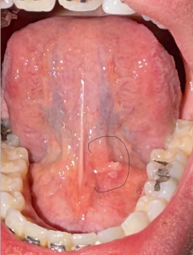
The photograph provides a clear view of the underside of the tongue, the lower anterior teeth, lower posterior teeth, and surrounding soft tissues.
The most obvious dental finding is a significant accumulation of hard yellow-brown material along the lingual surfaces of the lower anterior teeth. This appearance is strongly suggestive of mature dental calculus.
There are also areas of apparent gingival irritation and inflammation around the lower teeth.
The circled area beneath the tongue shows a localized raised or prominent soft-tissue structure. From this photograph alone, it cannot be determined whether this represents a normal anatomical structure, salivary-duct tissue, irritation, inflammation, trauma, cystic change, or another oral lesion.
It should therefore be examined directly rather than diagnosed from the photograph.
2. Preliminary Dental Assessment
Finding 1: Heavy Dental Calculus
The lower front teeth appear to have substantial calculus accumulation, particularly on the tongue-facing surfaces.
This is unlikely to be removed adequately through ordinary toothbrushing alone.
Professional scaling is indicated if clinical examination confirms the deposits are calculus.
Finding 2: Possible Gingival Inflammation
The surrounding gingiva appears irritated and somewhat red.
This may be consistent with plaque-induced gingivitis.
However, the photograph cannot determine whether the inflammation has progressed to periodontitis.
The critical measurements are periodontal pocket depth, clinical attachment loss, bleeding on probing, tooth mobility, recession, and radiographic bone levels.
Finding 3: Possible Periodontal Disease
Because the visible calculus burden is substantial, the patient should be evaluated for periodontal disease.
A dentist should specifically determine whether calculus exists below the gumline and whether periodontal pockets have developed.
If deep pockets and attachment or bone loss are present, ordinary preventive cleaning may not be sufficient.
Treatment could require periodontal scaling and root-surface instrumentation.
Finding 4: Circled Floor-of-Mouth Area
The circled area is clinically important.
The appearance could represent normal or relatively benign anatomy, particularly structures associated with the salivary ducts and glands, but the photograph is insufficient to establish this.
The dentist should examine:
-
Whether the area is soft or firm.
-
Whether it is painful.
-
Whether it changes size.
-
Whether saliva is expressed normally from the nearby duct.
-
Whether there is ulceration.
-
Whether there is bleeding.
-
Whether there is a white or red surface change.
-
Whether the lesion is fixed or movable.
-
Whether there are enlarged lymph nodes.
-
How long it has been present.
If the area is new, unexplained, enlarging, painful, ulcerated, or persistent, it should receive direct clinical assessment rather than simply being observed from photographs.
3. Recommended Diagnostic Process
Visit 1: Comprehensive Examination
The first appointment should include:
-
Full dental examination.
-
Periodontal charting.
-
Pocket-depth measurements.
-
Bleeding-on-probing assessment.
-
Tooth mobility assessment.
-
Evaluation of gingival recession.
-
Evaluation of calculus above and below the gumline.
-
Examination of the tongue and floor of mouth.
-
Evaluation of the circled lesion.
-
Radiographs when clinically indicated.
The purpose is to establish whether this is primarily gingivitis, periodontitis, or another condition requiring more extensive treatment.
4. Scaling and Periodontal Treatment
If examination confirms only superficial calculus and gingivitis, professional scaling and polishing may be appropriate.
If there is significant subgingival calculus and periodontal pocketing, the dentist may recommend scaling and root planing or other periodontal therapy.
Deep scaling is different from ordinary preventive cleaning because the dentist must access deposits below the gumline and clean affected root surfaces. Periodontal clinics in Cebu describe scaling and root planing as treatment for cases where plaque and tartar have progressed beyond simple gingivitis.
Treatment may be divided into several appointments depending on the amount of calculus, number of affected teeth, pocket depth, sensitivity, and overall periodontal condition.
5. Can All Teeth Be Scaled?
Potentially, yes, if professional examination confirms that full-mouth scaling is appropriate.
However, the dentist should not automatically perform aggressive scaling on every tooth simply because calculus is visible.
The treatment should be based on:
-
Amount of calculus.
-
Location of calculus.
-
Periodontal pocket depth.
-
Bone loss.
-
Gum inflammation.
-
Tooth mobility.
-
Root exposure.
-
Existing restorations.
-
Patient sensitivity.
-
Overall periodontal diagnosis.
A full-mouth treatment may require one appointment or multiple appointments.
6. Expected Healing Timeline
The healing period depends heavily on whether the patient has gingivitis or established periodontal disease.
First 24 to 72 hours
The gums may feel sensitive after significant calculus removal.
Minor bleeding can occur, particularly when inflamed tissue is being professionally cleaned.
The teeth may temporarily feel more sensitive.
Days 4 to 7
Inflammation should begin decreasing if plaque control is good and the irritant has been removed.
Gingival tissue may begin appearing healthier.
Days 7 to 14
A meaningful improvement in gingival inflammation may be visible.
Bleeding during brushing should decrease if the patient maintains effective oral hygiene.
The dentist can begin assessing the early response to treatment.
Around 2 to 6 weeks
A more meaningful periodontal reassessment may be appropriate, particularly when periodontal disease was suspected.
Pocket measurements and bleeding can be compared with the initial examination.
7. What If It Takes 14 Days?
Fourteen days is a reasonable period for observing improvement in gingival inflammation after professional treatment, but it should not be considered a guaranteed complete healing period.
The important question is not simply whether the gums look better after 14 days.
The dentist needs to determine whether:
-
Bleeding has decreased.
-
Swelling has decreased.
-
Gingival tissue has become healthier.
-
Periodontal pockets remain.
-
Calculus remains below the gumline.
-
Teeth remain mobile.
-
Bone loss is present.
-
The floor-of-mouth finding has changed.
8. What Could Scale Up If Treatment Is Delayed?
If the patient has periodontal disease and the condition remains uncontrolled, the disease may progress from inflammation to destruction of the supporting tissues around the teeth.
The potential progression is:
Plaque accumulation
Then calculus formation
Then persistent gingival inflammation
Then periodontal pocket formation
Then attachment loss
Then supporting bone loss
Then possible tooth mobility
Then possible tooth loss
However, this sequence is not inevitable for every patient. The actual risk depends on the patient's periodontal measurements, oral hygiene, smoking status, systemic factors, bacterial burden, and other clinical factors.
9. What Should Happen to the Circled Area?
This should be examined separately from the calculus problem.
Do not assume that the circled area is caused by tartar.
The dentist should inspect it during the same appointment.
If it represents normal anatomy or minor irritation, no major treatment may be necessary.
If it represents an inflammatory or salivary-gland-related problem, management will depend on the clinical findings.
If it is an unexplained persistent lesion, the dentist may recommend additional investigation or referral to an oral medicine or oral surgery specialist.
A lesion that persists or changes should not simply be ignored because the teeth have been cleaned.
10. Home Care During the 14-Day Period
After professional treatment, the patient should maintain excellent plaque control.
Recommended routine:
Morning:
Brush carefully with fluoride toothpaste.
Clean between the teeth.
Clean the tongue gently.
Evening:
Brush again for approximately two minutes.
Clean between the teeth.
Follow any mouthwash or medication instructions specifically provided by the dentist.
Avoid attempting to scrape hardened calculus from the teeth with household metal instruments.
11. When to Return Earlier
Do not wait 14 days if there is:
Rapidly increasing swelling.
Severe pain.
Pus.
Fever.
Difficulty swallowing.
Difficulty breathing.
Difficulty moving the tongue.
Rapid enlargement of the floor-of-mouth area.
Neck swelling.
Significant bleeding.
Rapidly worsening symptoms.
These findings require prompt professional assessment.
12. Finding a Dental Clinic in Cebu
For a Cebu-based patient, the most appropriate clinic should be selected based on the patient's actual location rather than simply choosing a clinic based on online ratings.
The Cebu dental directory can be used to search for a nearby clinic offering periodontal examination, scaling, and related dental services.
For example, current Cebu City listings include:
Near Dental Clinic - Mabolo
Affinity Dental Clinics Cebu
Cebu Dental Care Center is another Cebu City option with documented full-mouth scaling services. A recent patient review describes full-mouth scaling performed over two appointments.
CebuDENT Oral Care also lists both dental cleaning, scaling and polishing, and periodontal treatment among its services.
For the patient's actual nearest clinic, use the CebuDentalImplants.com directory location search and enter the patient's barangay, municipality, or city.
13. Recommended Case Classification
Based on the photograph alone:
Dental calculus: Significant
Gingival inflammation: Suspected
Periodontitis: Cannot be confirmed photographically
Subgingival calculus: Requires clinical examination
Bone loss: Cannot be determined from photograph
Floor-of-mouth lesion: Requires direct examination
Full-mouth scaling: Potentially indicated, depending on periodontal examination
14-day healing assessment: Reasonable for monitoring gingival response, but not a substitute for periodontal reassessment
Final Clinical Comment
The most obvious problem in this photograph is the substantial calculus accumulation around the lower teeth. The appropriate next step is not simply cosmetic cleaning. The patient should receive a complete periodontal assessment to determine whether the calculus has contributed to periodontal pocketing, attachment loss, or bone loss.
The floor-of-mouth area circled in the photograph should also be examined during the same appointment. It should not automatically be attributed to periodontal disease or calculus.
The most important objective is to establish the diagnosis first, remove the causative deposits professionally, control plaque, and reassess the tissues rather than assuming that everything will be completely healed within 14 days.
This photographic assessment should therefore be considered a preliminary case review, not a definitive diagnosis or treatment prescription.
Related Clinical Cases
Patients with lower front gum inflammation may also experience tartar buildup, periodontal disease, gum abscesses, or tooth decay. Explore these related clinical case studies.