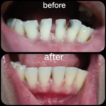
Strong Teeth, Fresh Breath, Brighter You.
Comprehensive Dental Case Analysis: Tooth Decay, Severity Grading, Treatment Plan & Healing Timeline
Image:
Severity:
Dental Case Full Analysis and Diagnosis
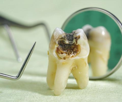
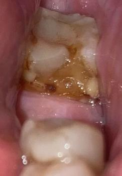
The clinical image shows a tooth with extensive decay. The enamel surface is disrupted and broken down, with dark discoloration indicating advanced carious involvement. This tooth likely exhibits significant decay reaching deeper tooth layers.
The working diagnosis is dental caries in an advanced stage. Further clinical evaluation is needed to determine whether the pulp is involved.
Severity Grading
| Severity Level | Features | Diagnosis |
|---|---|---|
| Mild | Early enamel demineralization, white spots | Not present |
| Moderate | Cavities visible, enamel and dentin involvement | Likely |
| Severe | Extensive decay, large cavity | Likely |
| Critical | Pulp infection risk, abscess possible | Not confirmed |
| Emergency | Severe pain or swelling | Not visible in image |
Assessment indicates moderate to severe dental caries. If the pulp is involved, the condition may be near critical.
Examination Considerations
A complete evaluation should include clinical exam, dental X-rays, and pulp vitality testing. These confirm decay depth and pulp involvement.
Expected Time Frame to Heal
Healing depends on the treatment performed:
- Restorative filling with no pulp involvement can be completed immediately, with sensitivity lasting up to two weeks.
- If pulp involvement exists a root canal and crown are recommended, with soft tissue healing in one to two weeks and full healing over weeks to months.
- Delaying treatment beyond 14 days increases risk of complications.
Risks If Treatment Is Delayed
Delaying care can result in:
- Spread of infection
- Increased pain
- Dental abscess
- Bone loss
- Potential extraction
Step-by-Step Treatment Plan
Step 1 Clinical and Radiographic Assessment
- Intraoral clinical evaluation
- Diagnostic X-rays
- Pulp vitality tests
Step 2 Infection Control
- Administer local anesthesia if needed
- Isolate the operative area
Step 3 Caries Removal
Remove decayed tissue and evaluate depth relative to pulp status.
Step 4 Definitive Treatment
If the pulp is not involved a restorative filling is placed. If the pulp is involved root canal therapy followed by a crown is recommended.
Step 5 Finishing and Polishing
Refine the restoration and ensure proper bite and comfort.
Step 6 Follow-Up
Check healing one to two weeks after treatment.
Comments and Clinical Interpretation
The decay is beyond early stages and likely involves deep tooth structures. Without prompt treatment the risk of infection and complications increases.
Recommended Next Step
Visit a dental clinic as soon as possible for a complete assessment and treatment. Use a dental directory to locate a provider in your area.
Search the directory for the nearest dental clinic: cebudentalimplants.com/map-dental-clinic
Patient Advice
Seek dental care promptly. Maintain oral hygiene with brushing and flossing. Avoid sticky or very cold foods. Seek treatment within seven to ten days where possible.
Teeth Case
 |
 |
 |
 |
 |
 |