Modern Dentistry, Timeless Smiles.

Why a Visual Dental Examination Alone Isn't Enough
image hero:
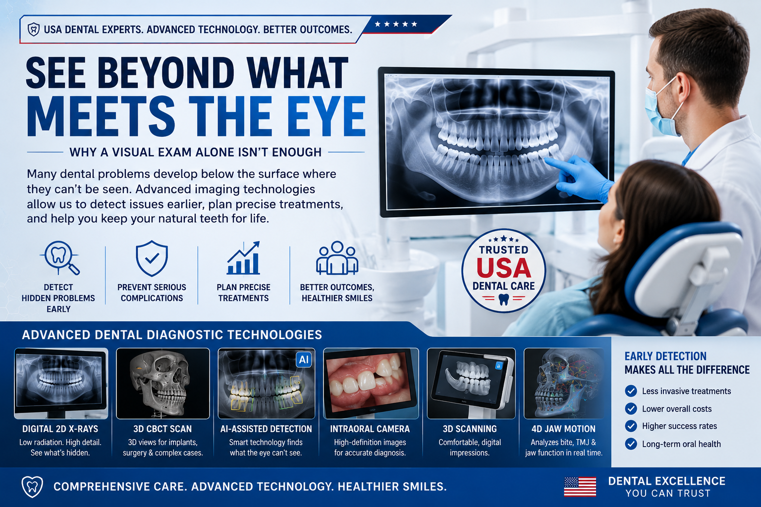
A USA Dentist Explains How Advanced Dental Imaging Detects Hidden Problems Before They Become Serious
A routine dental examination begins with a careful visual inspection of your teeth, gums, tongue, cheeks, and other oral tissues. Using a dental mirror, explorer, and specialized lighting, your dentist evaluates visible signs of decay, gum disease, fractures, worn restorations, and oral abnormalities. While this clinical examination is an essential first step, it only reveals what can be seen on the surface.
Modern dentistry recognizes that many of the most serious oral health conditions develop beneath the enamel, below the gumline, or deep within the jawbone—areas that cannot be evaluated through visual inspection alone. Hidden cavities, bone loss, root infections, impacted teeth, microscopic fractures, and early periodontal disease often remain completely invisible until they become painful or cause irreversible damage.
For this reason, leading dental practices throughout the United States combine comprehensive clinical examinations with advanced digital diagnostic technologies. These modern imaging systems allow dentists to identify disease at its earliest stages, preserve healthy tooth structure, reduce treatment complexity, and improve long-term outcomes.
Why a Visual Examination Alone Isn't Enough
Although a dentist can identify many visible oral health problems during an examination, several important limitations prevent a visual inspection from providing a complete diagnosis.
1. Hidden Tooth Surfaces Cannot Be Seen
A significant portion of every tooth is hidden from direct view.
These include:
- Areas between neighboring teeth (interproximal surfaces)
- Tooth roots beneath the gums
- Supporting jawbone
- Internal tooth structures
- Root canals
In fact, nearly half of a tooth's anatomy cannot be evaluated with the naked eye or a dental mirror alone.
Without diagnostic imaging, early disease may progress unnoticed for months or even years.
2. Early Tooth Decay Often Looks Completely Normal
One of the greatest challenges in dentistry is detecting cavities before they become large enough to require extensive treatment.
Early enamel demineralization frequently develops beneath an apparently healthy tooth surface.
A tooth may appear perfectly normal while microscopic decay continues beneath the enamel.
By the time discoloration or a visible hole appears, significantly more tooth structure may already be damaged.
Digital dental X-rays frequently reveal these hidden lesions long before they become visible during a routine examination.
3. Bone Loss Develops Below the Gumline
The jawbone that supports your teeth cannot be seen during a visual examination.
As periodontal disease progresses, bone destruction often occurs silently.
Patients frequently experience:
- no pain
- no obvious swelling
- no visible symptoms
until considerable bone has already been lost.
Dental imaging allows dentists to measure:
- bone height
- bone density
- bone defects
- periodontal attachment loss
allowing treatment before permanent tooth instability develops.
4. Root Infections Remain Hidden
A tooth may appear healthy above the gumline while infection is destroying bone around its root.
Conditions that commonly remain invisible include:
- chronic root infections
- periapical abscesses
- cyst formation
- internal root resorption
- external root resorption
These conditions are usually diagnosed only through radiographic imaging.
5. Problems Under Existing Dental Restorations
Even well-made crowns and fillings can eventually develop recurrent decay.
Because these areas are covered by restorative materials, dentists often cannot detect secondary decay during a visual examination.
- hidden cavities beneath fillings
- leaking crown margins
- failing bridge abutments
- recurrent decay beneath restorations
before complete restoration failure occurs.
Advanced Dental Diagnostic Technologies Used in the United States
Modern American dental practices combine clinical expertise with sophisticated digital imaging technologies that evaluate oral structures far beyond what the eye can see.
Digital Dental Radiography
Digital dental X-rays provide high-resolution images of:
- teeth
- roots
- supporting bone
- periodontal structures
- hidden cavities
Benefits
- Detects decay between teeth
- Reveals bone loss
- Identifies root infections
- Evaluates previous dental work
- Produces images within seconds
- Uses significantly less radiation than traditional film systems
3D Cone Beam Computed Tomography (CBCT)
CBCT technology produces three-dimensional images of the entire oral and maxillofacial region.
Unlike conventional dental X-rays, CBCT allows dentists to examine structures from every angle.
Common Uses
- Dental implant planning
- Wisdom tooth evaluation
- Root canal anatomy
- Jawbone assessment
- Sinus evaluation
- Nerve mapping
- Oral surgery planning
- TMJ evaluation
CBCT has become the gold standard for complex dental diagnosis and surgical planning.
AI-Assisted Dental Imaging
Artificial intelligence is becoming an important tool in modern diagnostic dentistry.
AI software analyzes digital X-rays using advanced machine-learning algorithms that highlight subtle abnormalities that might otherwise be overlooked.
AI-assisted analysis helps identify:
- microscopic cavities
- enamel demineralization
- bone height changes
- early periodontal disease
- suspicious radiographic findings
AI supports clinical decision-making but does not replace the expertise of a licensed dentist.
Intraoral Cameras and Digital 3D Scanners
High-definition intraoral cameras provide magnified views of teeth and soft tissues.
Digital scanners create precise three-dimensional models without traditional impression materials.
These technologies help dentists identify:
- tiny enamel cracks
- worn restorations
- defective crown margins
- tooth wear
- erosion
- cosmetic concerns
Patients can also view these images in real time, making it easier to understand their diagnosis and treatment options.
Near-Infrared Transillumination (NIRI)
Near-infrared imaging uses light rather than ionizing radiation to evaluate tooth structure.
This technology is especially useful for detecting:
- early interproximal decay
- enamel cracks
- hidden lesions
- structural defects
Because no X-rays are used, NIRI can be an excellent adjunctive tool for ongoing monitoring in appropriate clinical situations.
Digital Occlusal Analysis and Jaw Motion Technology
Modern digital bite analysis systems evaluate how teeth come together during function.
Advanced jaw tracking technology records:
- chewing patterns
- bite force
- jaw movement
- TMJ function
- occlusal imbalance
These dynamic assessments provide information that cannot be obtained from a static visual examination alone.
Why Combining Technologies Produces Better Diagnoses
No single diagnostic method can identify every dental problem.
The most accurate diagnoses come from combining:
- Comprehensive clinical examination
- Digital dental X-rays
- 3D CBCT imaging (when indicated)
- Intraoral photography
- Digital scanning
- AI-assisted image analysis
- Periodontal evaluation
- Functional bite assessment
Together, these technologies provide a complete picture of your oral health, allowing dentists to detect disease earlier, recommend less invasive treatments, improve long-term success rates, and help preserve your natural teeth for years to come.
Key Takeaway
A visual dental examination is an essential part of every checkup, but it tells only part of the story. Many serious conditions—including hidden cavities, root infections, bone loss, periodontal disease, and jaw abnormalities—develop beneath the surface long before symptoms appear. By combining clinical expertise with advanced diagnostic technologies such as digital radiography, 3D CBCT, AI-assisted imaging, intraoral scanners, and near-infrared detection systems, dentists can identify problems earlier, provide more precise treatment planning, and help patients maintain healthier smiles with fewer complications over time.
Dentist Hub article to:
- What Dental X-Rays Reveal
- Why Dentists Use Dental X-Rays
- Digital Dental X-Rays
- CBCT (Cone Beam Computed Tomography)
- Clinical Examination
- Interproximal Tooth Decay
- Hidden Tooth Infection
- Jawbone Loss
- Periodontal Disease
- Dental Implant Planning
- TMJ Disorders
- Intraoral Scanner
- AI in Dentistry
- Dental X-Ray Radiation and Safety
Related Clinical Cases
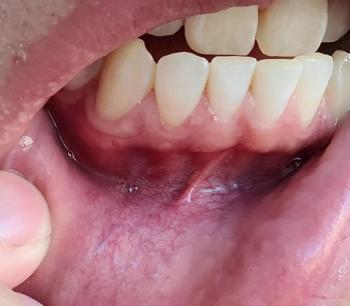
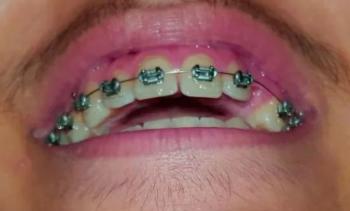
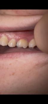
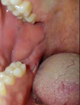
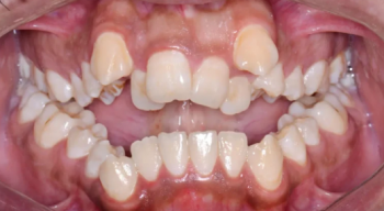
Patients with lower front gum inflammation may also experience tartar buildup, periodontal disease, gum abscesses, or tooth decay. Explore these related clinical case studies.



Other topics:
Other Dental Clinic