Your Smile, Perfected with Precision.

How Dental X-Rays Prevent Misdiagnosis and Treatment Errors: A USA Dentist Explains the Technology Behind Accurate Dental Care
image hero:
Reviewed by a USA Dentist | Digital Dentistry & Diagnostic Imaging
Modern dentistry is built on one fundamental principle: accurate diagnosis leads to successful treatment. No matter how experienced a dentist may be, many serious dental conditions develop beneath the surface of the teeth and gums where they cannot be seen during a routine clinical examination.
Dental X-rays (radiographs) provide objective, measurable, and scientifically validated images of internal oral structures that remain completely invisible to the naked eye. Without radiographic imaging, clinicians would have to rely primarily on visual inspection and patient symptoms, significantly increasing the risk of diagnostic errors, inappropriate treatment planning, delayed care, or unnecessary procedures.
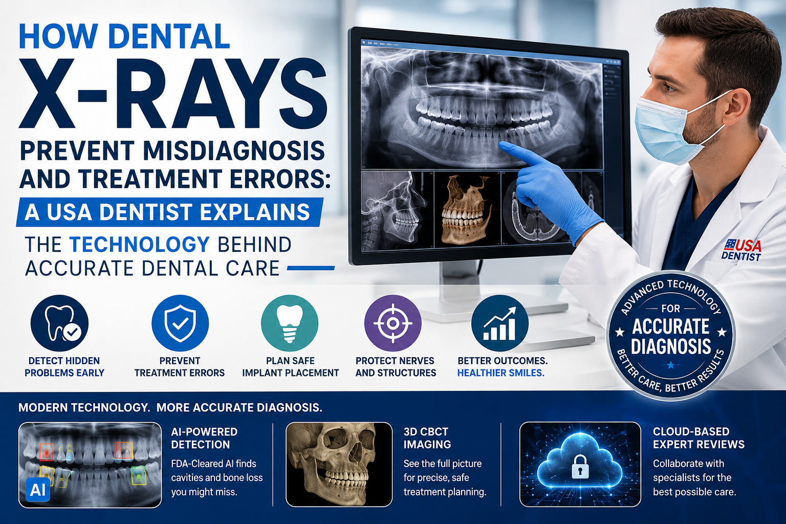
Today's digital imaging technologies—including FDA-cleared artificial intelligence (AI), Cone Beam Computed Tomography (CBCT), and cloud-based diagnostic collaboration platforms—have transformed dental diagnosis by reducing human error while improving precision and patient safety.
Why Visual Examination Alone Is Not Enough
During a comprehensive oral examination, dentists carefully evaluate:
- Teeth
- Gums
- Bite alignment
- Oral tissues
- Tongue
- Existing restorations
However, a clinical examination cannot see:
- Tooth roots
- Jawbone
- Bone density
- Hidden cavities
- Internal root canals
- Impacted teeth
- Nerve pathways
- Developing infections beneath the gums
Many serious dental diseases progress silently before becoming painful. By the time symptoms appear, treatment often becomes more extensive and costly.
Digital radiographs allow dentists to identify disease at its earliest stages, when treatment is typically simpler, less invasive, and more predictable.
How Dental X-Rays Prevent Diagnostic Errors
1. Preventing Root Canal Treatment Failure
One of the most common reasons root canal therapy fails is untreated anatomy.
Many teeth contain:
- Hidden accessory canals
- Extremely narrow canals
- Curved canals
- Extra roots
- Calcified canals
These structures cannot be identified through visual examination alone.
Digital periapical radiographs and CBCT imaging reveal the complete internal anatomy of the tooth, allowing the dentist to clean, disinfect, and seal every canal.
Without X-rays, untreated canals may continue harboring bacteria, resulting in:
- Persistent infection
- Chronic pain
- Dental abscess
- Retreatment
- Tooth extraction
Radiographic imaging greatly improves long-term root canal success.
2. Preventing Injury to the Inferior Alveolar Nerve
Lower wisdom teeth and dental implants are often located close to the inferior alveolar nerve, which supplies sensation to the:
- Lower lip
- Chin
- Lower teeth
Without precise imaging, surgery may unintentionally damage this nerve.
Potential complications include:
- Permanent numbness
- Tingling sensations
- Altered facial sensation
- Difficulty speaking
- Difficulty eating
Panoramic X-rays provide an overall view of the jaws, while CBCT scans display the nerve's exact three-dimensional location.
This allows surgeons to safely plan procedures while minimizing neurological complications.
3. Preventing Incorrect Dental Implant Placement
Dental implants require sufficient:
- Bone height
- Bone width
- Bone density
- Safe anatomical clearance
Visual examination cannot accurately measure these structures.
Before implant surgery, digital imaging evaluates:
- Available bone volume
- Maxillary sinus location
- Inferior alveolar nerve position
- Adjacent tooth roots
- Bone quality
Without radiographic planning, implants may be placed into:
- Sinus cavities
- Thin bone
- Weak bone
- Adjacent roots
- Nerve canals
This increases the risk of implant instability, surgical complications, nerve injury, and implant failure.
Modern implant dentistry relies heavily on digital imaging for predictable outcomes.
4. Detecting Hidden Cavities Before They Become Major Problems
Many cavities begin where the human eye cannot see.
Examples include:
- Between adjacent teeth
- Under crowns
- Beneath fillings
- Around bridges
- Along restoration margins
These lesions often remain invisible until extensive tooth destruction occurs.
Digital bitewing radiographs detect early decay long before symptoms develop.
Early diagnosis allows dentists to:
- Preserve healthy tooth structure
- Use smaller restorations
- Avoid root canal therapy
- Prevent tooth loss
The earlier decay is identified, the more conservative treatment can be.
5. Preventing Overtreatment and Undertreatment
Accurate diagnosis is not only about finding disease—it is also about determining whether treatment is truly necessary.
Dental X-rays help distinguish:
Early Enamel Demineralization
This stage often responds to:
- Fluoride therapy
- Improved oral hygiene
- Dietary modification
- Remineralization treatments
No drilling may be necessary.
Deep Dentin Decay
When decay extends into dentin, restorative treatment becomes necessary to prevent pulp infection.
Radiographs allow dentists to objectively determine which lesions require treatment and which can be monitored conservatively.
This prevents both:
- Unnecessary drilling (overtreatment)
- Delayed intervention (undertreatment)
Additional Diagnostic Errors Prevented by Dental X-Rays
Digital radiography also helps detect conditions that may otherwise remain hidden, including:
- Vertical root fractures
- Internal root resorption
- External root resorption
- Bone loss from periodontal disease
- Impacted teeth
- Cysts
- Jaw tumors
- Chronic dental abscesses
- Sinus-related dental complications
- Failed previous dental treatments
Many of these conditions produce little or no discomfort during their early stages.
Modern Technologies Reducing Human Diagnostic Error
Dentistry in the United States increasingly integrates advanced technologies that improve diagnostic consistency and treatment planning.
FDA-Cleared Artificial Intelligence (AI) Diagnostic Software
Several FDA-cleared AI systems, including platforms such as Overjet and Align X-ray Insights, assist dentists by analyzing digital radiographs in seconds.
These systems can:
- Highlight suspected cavities
- Measure periodontal bone loss
- Identify calculus deposits
- Detect radiographic abnormalities
- Standardize measurements
- Improve communication with patients
Clinical research has shown that AI-assisted interpretation can improve the detection of interproximal caries compared with unaided visual review, helping clinicians identify subtle lesions that may otherwise be overlooked.
Importantly, AI does not replace the dentist. Instead, it serves as a decision-support tool that enhances diagnostic accuracy while the dentist remains responsible for the final diagnosis and treatment plan.
Cone Beam Computed Tomography (CBCT)
Traditional dental X-rays create two-dimensional images.
CBCT technology captures a complete three-dimensional representation of the patient's oral anatomy.
Advantages include:
- Elimination of overlapping structures
- Precise bone measurements
- Accurate implant planning
- Detailed root canal anatomy
- Identification of hidden pathology
- Improved surgical planning
CBCT has become the standard of care for many complex procedures, including dental implants, impacted wisdom teeth, endodontics, and oral surgery.
Cloud-Based Digital Consultation
Digital dentistry also enables secure collaboration among multiple dental professionals.
Cloud-based imaging platforms allow:
- Remote specialist consultation
- Second opinions
- Orthodontic planning
- Oral surgery referrals
- Implant treatment planning
- Endodontic case review
Because high-resolution digital radiographs can be shared instantly, specialists can evaluate the same images regardless of geographic location.
This collaborative approach reduces diagnostic variability and supports more consistent treatment decisions.
Benefits for Patients
Modern digital radiography provides significant advantages beyond diagnosis.
Patients benefit from:
- Earlier disease detection
- More accurate treatment planning
- Fewer unexpected complications
- Greater preservation of healthy teeth
- Reduced need for invasive procedures
- Improved long-term treatment success
- Better understanding of their oral health through visual imaging
Digital X-rays also expose patients to significantly less radiation than older film-based systems while producing higher-quality diagnostic images.
Frequently Asked Questions
Can a dentist diagnose everything without X-rays?
No. Many important dental conditions—including cavities between teeth, root infections, bone loss, impacted teeth, and hidden fractures—cannot be detected through visual examination alone.
Are dental X-rays safe?
Yes. Modern digital dental X-rays use very low radiation doses and follow the ALARA principle (As Low As Reasonably Achievable). Dentists prescribe radiographs only when they are clinically necessary.
Why are CBCT scans sometimes recommended instead of regular X-rays?
CBCT provides three-dimensional images that reveal anatomical details not visible on standard two-dimensional radiographs. They are especially valuable for dental implants, complex root canal treatment, impacted teeth, and oral surgery planning.
Does artificial intelligence replace my dentist?
No. AI serves as an additional diagnostic aid that helps identify subtle findings and standardize measurements. Your dentist evaluates the clinical examination, radiographs, symptoms, and medical history before making the final diagnosis and treatment recommendations.
The Bottom Line
Dental X-rays are one of the most important tools in modern dentistry because they reveal disease hidden beneath the surface and reduce the risk of misdiagnosis. By accurately visualizing tooth roots, bone, nerve pathways, and early decay, radiographs help dentists plan safer, more predictable treatments while avoiding unnecessary procedures.
Today's digital innovations—including FDA-cleared AI-assisted image analysis, high-resolution CBCT imaging, and cloud-based specialist collaboration—continue to improve diagnostic accuracy and patient safety. Rather than replacing clinical judgment, these technologies provide dentists with more complete information, leading to better-informed decisions and improved long-term oral health outcomes.
Related Clinical Cases
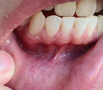
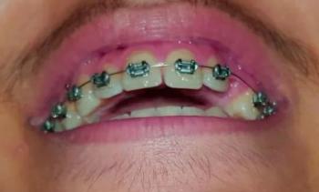
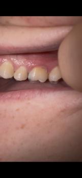
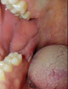
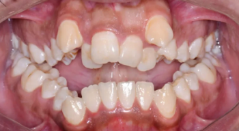
Patients with lower front gum inflammation may also experience tartar buildup, periodontal disease, gum abscesses, or tooth decay. Explore these related clinical case studies.



Other topics:
Other Dental Clinic