Because Every Tooth Deserves Care.

Bone Loss Around Front Teeth With Braces X-Ray Case
Image:

Severity:
Bone Loss Around Front Teeth With Braces X-Ray Case Analysis
What Is Seen in This Case
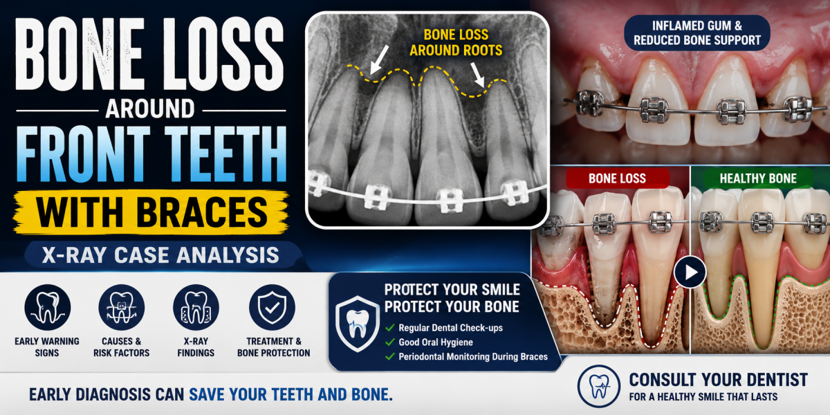
The X-ray image shows the lower front teeth with orthodontic braces in place. The tooth roots are visible, but the bone level around the roots appears reduced and uneven. There are darker areas between and around the roots, suggesting loss of supporting bone rather than healthy, dense bone.
This finding is especially important in patients undergoing orthodontic treatment.
Most Likely Diagnosis
Based on the X-ray appearance, the most likely conditions include:
-
Early to moderate periodontal bone loss
-
Bone resorption around front teeth
-
Gum disease progressing during orthodontic treatment
-
Inflammation-related bone changes
-
Increased risk of tooth mobility
This is not a cavity issue, but a supporting bone problem.
Why Bone Loss Can Occur With Braces
Common contributing factors include:
-
Plaque buildup around brackets and wires
-
Difficulty cleaning around orthodontic appliances
-
Pre-existing gum disease before braces
-
Excessive orthodontic force
-
Poor flossing and interdental cleaning
Braces can accelerate bone loss if gum health is not well controlled.
Is This a Serious Problem
Yes. Bone loss around front teeth is progressive if untreated. If ignored, it may scale up into:
-
Increased tooth mobility
-
Gum recession
-
Long-term instability after braces
-
Early tooth loss
-
Need to stop or modify orthodontic treatment
Early detection helps preserve teeth.
Bone Loss in Front Teeth: Expert Guide to X-Ray Findings, Braces, and Treatment Options
Understanding Bone Loss Around the Front Teeth
Bone loss around the front teeth occurs when the alveolar bone—the bone that supports the tooth roots—becomes reduced or damaged. This can affect the upper or lower incisors and, in advanced cases, may cause gum recession, tooth mobility, changes in tooth position, and eventual tooth loss.
When bone loss appears on a dental X-ray, the finding should be evaluated alongside a clinical examination. An X-ray can show the level and pattern of supporting bone, but it cannot determine the complete periodontal diagnosis on its own.
From a periodontal perspective, the first priority is to identify the cause of bone loss, determine whether the condition is active, and establish whether the affected teeth can be maintained safely.
What Causes Bone Loss in Front Teeth?
Several conditions can contribute to bone loss around the anterior teeth.
1. Periodontitis
Periodontitis is an inflammatory disease associated with dental plaque biofilm that damages the supporting tissues around teeth. As the disease progresses, the periodontal ligament and alveolar bone can be destroyed.
Common warning signs include:
-
Bleeding or swollen gums
-
Persistent bad breath
-
Gum recession that makes teeth appear longer
-
Deep periodontal pockets
-
Loose or drifting front teeth
Smoking, poorly controlled diabetes, inadequate plaque control, and individual susceptibility can increase the risk or severity of periodontal disease.
2. Orthodontic treatment and braces
Braces do not automatically cause bone loss. However, tooth movement can increase periodontal risks when significant inflammation is present, oral hygiene is inadequate, or teeth are moved beyond the limits of their supporting bone.
Patients with existing bone loss may need modified orthodontic mechanics, lighter or carefully controlled forces, and closer monitoring by an orthodontist and periodontist.
Important: Active periodontitis should be treated and brought under control before elective orthodontic tooth movement proceeds. Patients already wearing braces who develop progressive bone loss need prompt reassessment of their periodontal condition and orthodontic treatment plan.
3. Traumatic forces and tooth-position problems
Excessive biting forces, traumatic contact between teeth, or certain tooth-position abnormalities can contribute to mobility or complicate periodontal management. These factors should be assessed clinically rather than assumed to be the primary cause of bone loss.
4. Local dental infection or other pathology
A tooth with a history of trauma, root damage, or infection may develop localized bone changes. Less commonly, other conditions can resemble periodontal bone loss on an X-ray.
The dentist must distinguish these possibilities because treatment for an endodontic infection, a periodontal defect, and a combined endodontic-periodontal lesion can be different.
How Dentists Evaluate Bone Loss on an X-Ray
A periapical dental X-ray is often useful for evaluating the roots and surrounding bone of the front teeth. The dentist assesses the height of the alveolar crest, the pattern of bone loss, root condition, and any signs of localized pathology.
The examination should also include:
-
Periodontal probing to measure pocket depths
-
Bleeding on probing and gum-recession assessment
-
Evaluation of tooth mobility and tooth migration
-
Review of previous X-rays, when available, to assess changes over time
-
Medical and dental history, including smoking, diabetes, trauma, and orthodontic treatment
Bone loss may be described as horizontal or vertical. Horizontal loss generally involves a more even reduction in bone height, whereas vertical or angular defects extend unevenly alongside a tooth root.
These patterns help guide treatment planning, but the appearance of a single X-ray cannot reliably establish whether the disease is currently progressing. Clinical findings and comparison with earlier records are important.
Bone Loss Treatment: What Can Be Done?
Treatment depends on the cause, severity, remaining bone support, tooth mobility, and the patient's overall periodontal health.
1. Professional periodontal cleaning
For patients with plaque and calculus accumulation, professional cleaning removes deposits that cannot be adequately removed with routine brushing.
When periodontitis is diagnosed, scaling and root planing may be recommended. This procedure cleans the root surfaces below the gumline to reduce bacterial deposits and periodontal inflammation.
Cleaning can help control the disease, but it does not automatically rebuild bone that has already been lost.
2. Control of periodontal risk factors
Long-term success requires consistent plaque control and management of contributing factors. Patients should brush twice daily with fluoride toothpaste, clean between their teeth daily, stop tobacco use, and work with their healthcare provider to manage diabetes when applicable.
The dentist should reassess the response to treatment rather than assume the infection has resolved simply because bleeding or discomfort has decreased.
3. Periodontal surgery
Some patients with persistent deep pockets or inaccessible root surfaces may benefit from periodontal surgery. Depending on the defect, a periodontist may recommend access-flap surgery, pocket-reduction procedures, or other site-specific treatment.
Surgery is not necessary for every patient with radiographic bone loss. The decision depends on the clinical response to initial treatment and the anatomy of the affected area.
4. Bone grafting and periodontal regeneration
In selected cases, regenerative periodontal surgery may be considered. Depending on the type of bone defect and the condition of the surrounding tissues, a clinician may use bone-grafting materials, barrier membranes, or biologically active materials to support regeneration.
Not every bone defect is suitable for regeneration. A graft does not guarantee complete bone restoration, and the procedure may not be appropriate when infection is uncontrolled or the remaining tooth support is inadequate.
5. Orthodontic treatment after periodontal stabilization
Patients who need braces and already have reduced bone support may still be candidates for orthodontic treatment. However, the periodontal condition should first be stabilized, and the proposed tooth movements must be compatible with the remaining supporting tissues.
A coordinated plan between the orthodontist and periodontist can help reduce the risk of further damage. During treatment, periodontal monitoring and excellent oral hygiene are essential.
6. Extraction and replacement when a tooth cannot be maintained
Extraction may be considered if a tooth has an extremely poor prognosis because of severe attachment loss, uncontrolled infection, or other structural problems. It should not be recommended solely because an X-ray shows some bone loss.
When a front tooth cannot be saved, replacement options may include a dental implant, a fixed dental bridge, or a removable partial denture. Implant placement requires separate assessment of bone volume, gum health, bite forces, and overall suitability.
Can Lost Bone Around Front Teeth Grow Back?
The answer depends on the type and extent of the defect.
Once periodontitis has destroyed supporting bone, routine cleaning and home care generally do not restore the lost bone to its original level. They are nevertheless essential because controlling the disease helps prevent additional destruction.
Selected defects may respond to periodontal regenerative treatment. The amount of regeneration varies, and complete restoration cannot be promised.
Gum grafting is a separate procedure that primarily addresses soft-tissue deficiencies, such as recession. It does not automatically restore the underlying alveolar bone.
When Should You See a Dentist?
Arrange a periodontal assessment if an X-ray shows bone loss around the front teeth, particularly if you have bleeding gums, increasing tooth mobility, gum recession, or teeth that appear to be moving apart.
Seek urgent dental assessment if you develop significant swelling, pus, rapidly worsening pain, or a sudden change in tooth mobility. Facial swelling accompanied by fever or spreading redness requires prompt medical or dental care. Difficulty swallowing or breathing, or swelling extending toward the eye or neck, requires emergency care.
Expert Clinical Takeaway
The most important objective in treating bone loss around the front teeth is to control the underlying disease and preserve teeth that have a reasonable long-term prognosis.
An X-ray finding is the beginning of the diagnostic process, not the final diagnosis. Accurate treatment planning combines radiographic findings, periodontal measurements, tooth stability, risk factors, and the patient's treatment goals.
With appropriate periodontal treatment, good home care, and ongoing maintenance, many patients can keep their natural front teeth even when some supporting bone has already been lost.
Frequently Asked Questions
1. Is bone loss around the front teeth serious?
It can be serious because the bone supports the roots of the teeth. The significance depends on the amount and pattern of bone loss, whether the condition is progressing, and whether the teeth remain stable. Early assessment helps identify the most appropriate treatment.
2. Can braces make existing bone loss worse?
They can increase risk when orthodontic forces are applied in the presence of active periodontal disease or when teeth are moved beyond the limits of their supporting bone. Braces are not automatically harmful to every patient with reduced bone support. Periodontal stabilization and coordinated monitoring are essential.
3. Should I remove my braces if my X-ray shows bone loss?
Do not make that decision based on the X-ray alone. Contact your orthodontist and request a periodontal evaluation. If active disease or progressive damage is identified, the orthodontist may need to pause or modify active tooth movement while periodontal treatment is provided.
4. Can deep cleaning stop bone loss?
Scaling and root planing can reduce periodontal inflammation and help control periodontitis. Whether disease progression stops depends on treatment response, home care, risk factors, and maintenance. Deep cleaning does not guarantee that all previously lost bone will return.
5. Is bone grafting always necessary?
No. Many patients can be managed with nonsurgical periodontal treatment and regular maintenance. Regenerative surgery is considered only when the clinical findings and bone-defect anatomy make it appropriate.
6. Can a loose front tooth become firm again?
Sometimes. If mobility is partly caused by inflammation, the tooth may become more stable after periodontal treatment. However, extensive loss of supporting attachment may leave persistent mobility. The prognosis depends on the remaining support and other contributing factors.
7. Does gum recession mean I have bone loss?
Not necessarily. Gum recession means the gum margin has moved away from its original position. It can occur with or without significant periodontal bone loss. A clinical examination and appropriate imaging are needed to distinguish the causes.
8. Can bone loss around front teeth be reversed naturally?
Good oral hygiene can reduce inflammation and help prevent further damage, but it does not reliably regenerate bone already destroyed by periodontitis. Selected defects may be candidates for professional regenerative treatment.
9. How often should I have my gums checked after periodontal treatment?
The maintenance interval should be individualized according to disease severity, treatment response, oral hygiene, and risk factors. Some patients need professional periodontal maintenance every three to four months, while others may need a different interval determined by their dentist or periodontist.
10. Can a dental implant replace a front tooth lost because of bone loss?
Potentially, yes. However, the infection must be controlled, and the dentist must evaluate the available bone, gum tissue, bite, and overall health. Bone grafting may be needed in some cases, and an implant is not automatically the best option for every patient.
Medical disclaimer: This article provides general dental education and does not replace an in-person examination, diagnosis, or individualized treatment plan from a licensed dentist or periodontist.
Recommended Treatment Process
Initial Assessment (Days 1–3)
-
Full periodontal examination
-
Review of orthodontic forces
-
Comparison with previous X-rays
-
Gum pocket measurements
Active Treatment Phase (Days 4–10)
Treatment may include:
-
Professional deep cleaning
-
Improved oral hygiene protocol
-
Adjustment of orthodontic force if needed
-
Antibacterial mouth rinse
-
Periodontal monitoring
Healing and Stabilization Phase (Days 11–14)
-
Reduced inflammation
-
Slowing of bone loss
-
Stabilization of tooth support
Long-term monitoring is essential.
Expected Healing Time
-
Gum inflammation reduction: 7–14 days
-
Bone stabilization: weeks to months
-
Bone loss already present does not fully regenerate
Early management prevents further damage.
What Happens If Treatment Is Delayed
Delaying care may lead to:
-
Continued bone destruction
-
Loose front teeth
-
Poor orthodontic outcome
-
Permanent gum recession
-
Premature tooth loss
This condition will not self-correct.
Home Care While Wearing Braces
Patients should:
-
Brush after every meal
-
Use interdental brushes around brackets
-
Floss daily with orthodontic floss
-
Rinse with dentist-recommended mouthwash
-
Attend regular cleaning appointments
Good hygiene is critical during braces.
Professional Comment
This X-ray shows bone loss around lower front teeth during orthodontic treatment, a condition that requires close coordination between dentist and orthodontist. Early intervention can preserve tooth support and allow safe continuation of braces.
Expert Dental Guidance: Bone Loss Around the Front Teeth During Orthodontic Treatment
Bone loss around the front teeth can have several causes, and an X-ray finding should not automatically be interpreted as being caused by braces. Orthodontic tooth movement, existing gum disease, thin bone around a tooth, previous trauma, tooth position, and other periodontal conditions can all influence the amount of supporting bone visible around the teeth.
A dentist or periodontist should evaluate the clinical examination together with dental X-rays before determining the cause and appropriate treatment.
What Does Bone Loss Around a Front Tooth Mean?
The teeth are supported by the gums, periodontal ligament, and alveolar bone. When supporting bone is lost, the tooth may have less structural support.
Bone loss may appear as:
-
Reduced bone height around the tooth
-
A localized vertical or angular bone defect
-
Thinning of the bone on the front or back surface of a tooth
-
Increased periodontal probing depth
-
Gum recession with an exposed root surface
-
Tooth mobility in more advanced cases
An X-ray can provide important information, but it does not tell the entire story. Some defects—particularly thin areas of bone on the facial side of a tooth—may be difficult to evaluate accurately on conventional two-dimensional X-rays.
Can Braces Cause Bone Loss?
Orthodontic treatment does not automatically cause permanent bone loss.
When braces move teeth, the supporting tissues undergo normal remodeling. Bone is removed on one side of a tooth and deposited on the other side as the tooth changes position.
However, excessive or unfavorable tooth movement, pre-existing periodontal disease, poor oral hygiene, or movement beyond the limits of the available supporting bone may increase the risk of periodontal complications in susceptible patients.
This is why orthodontic treatment should be planned according to the patient's periodontal health, tooth position, bone anatomy, and individual risk factors.
What If Bone Loss Was Already Present Before Braces?
This is an important distinction.
If bone loss existed before orthodontic treatment, the braces may not be the original cause. A patient may have had:
-
Periodontitis
-
Previous gum inflammation
-
Gum recession
-
A thin facial bone plate
-
Previous dental trauma
-
A tooth positioned outside the ideal bone envelope
-
Previous orthodontic treatment
-
Localized periodontal defects
A dentist should compare previous and current records whenever possible to determine whether the condition is stable, improving, or progressing.
What Should a Dentist Check?
A comprehensive evaluation may include:
-
Periodontal probing to measure gum pocket depths.
-
Assessment of bleeding and inflammation.
-
Evaluation of gum recession.
-
Checking tooth mobility.
-
Review of dental X-rays.
-
Comparison with previous X-rays when available.
-
Evaluation of tooth position and orthodontic movement.
-
Assessment of oral hygiene and plaque/calculus accumulation.
-
Periodontal examination before continuing or modifying orthodontic treatment.
In selected cases, a dentist or specialist may recommend additional three-dimensional imaging when the information is clinically necessary. CBCT should not be performed routinely simply because a patient is concerned about bone loss; the benefits and radiation exposure should be considered.
Dentist Expert Voice
Dentist's perspective: “An X-ray showing reduced bone support around a front tooth should be interpreted in the context of the patient's periodontal examination and orthodontic history. The key question is not simply whether bone loss is present, but why it is present, whether it is active, and whether the tooth remains adequately supported.”
A periodontist may be particularly helpful when there is significant bone loss, persistent gum inflammation, recession, deep periodontal pockets, or tooth mobility.
An orthodontist can assess whether the current tooth position and planned tooth movement are appropriate for the available supporting tissues.
In some cases, collaboration between the orthodontist and periodontist provides the most appropriate approach.
Does Bone Grow Back After Orthodontic Treatment?
Bone does not necessarily return to its original shape simply because orthodontic treatment is stopped.
The outcome depends on the cause, severity, location, periodontal health, and whether the underlying disease is controlled.
If active periodontal disease is present, periodontal treatment and control of inflammation are important before assuming that orthodontic treatment alone will solve the problem.
Some periodontal defects may respond to periodontal therapy, while selected defects may require additional periodontal procedures. Treatment must be individualized after examination.
When Should You See a Dentist or Periodontist?
Arrange a dental evaluation if you notice:
-
Increasing tooth mobility
-
Persistent bleeding when brushing or flossing
-
Gum recession
-
Swelling or tenderness around a tooth
-
Persistent bad breath
-
Deep periodontal pockets
-
Increasing sensitivity around a receding gum
-
A noticeable change in tooth position
-
A new or worsening gap between teeth
-
An X-ray showing significant supporting-bone loss
Seek prompt professional evaluation if there is rapid swelling, pus, severe pain, fever, facial swelling, or difficulty swallowing or breathing, because these symptoms can indicate a more serious dental infection.
Visit a Dental Clinic Near You
For periodontal evaluation during orthodontic treatment, visit:
https://cebudentalimplants.com/map-dental-clinic
Bone loss around the front teeth is a serious dental condition that can threaten the stability of your smile if left untreated. In this video, we analyze a real clinical case using a dental X-ray to explain how bone loss develops around the front teeth, especially in patients wearing braces or with untreated gum disease.
You'll learn what dentists look for on an X-ray, the common causes of alveolar bone loss, early warning signs, treatment options, and whether the damage can be stopped or managed before tooth loss occurs.
In this video you'll discover:
- What bone loss around front teeth looks like on a dental X-ray
- Can braces contribute to bone loss?
- How gum disease affects the jawbone
- Early symptoms of periodontal bone loss
- Treatment options to protect your teeth
- How dentists monitor bone health during orthodontic treatment
- Ways to prevent further bone loss
Frequently Asked Questions (FAQ): Bone Loss Around Front Teeth With Braces – X-Ray Findings, Causes, Treatment, and Prevention
1. What does bone loss around the front teeth mean?
Bone loss around the front teeth occurs when the jawbone that supports the roots begins to shrink due to periodontal (gum) disease, chronic inflammation, trauma, or other underlying conditions. As the supporting bone decreases, the teeth may become loose, shift position, and eventually be at risk of tooth loss if treatment is delayed.
2. Can braces cause bone loss around the front teeth?
Braces themselves do not cause bone loss when orthodontic treatment is properly planned and monitored. However, excessive orthodontic forces, poor oral hygiene, untreated gum disease, or pre-existing periodontal problems can increase the risk of bone loss during treatment.
3. How is bone loss detected on a dental X-ray?
Dentists evaluate bone height surrounding the tooth roots using dental X-rays such as:
- Periapical X-rays
- Bitewing X-rays
- Panoramic X-rays
- Cone Beam CT (CBCT)
Reduced bone height, widened periodontal ligament spaces, and changes in bone density are common radiographic signs.
4. What are the early signs of bone loss around the front teeth?
Early symptoms may include:
- Bleeding gums
- Swollen or red gums
- Gum recession
- Persistent bad breath
- Tooth sensitivity
- Slight tooth movement
- Spaces developing between front teeth
Many patients experience no pain during the early stages.
5. Can bone loss around front teeth be reversed?
Lost bone generally does not regenerate on its own. However, early treatment can stop further destruction. In selected cases, regenerative procedures such as bone grafting, guided tissue regeneration (GTR), or biologic materials may help restore some supporting bone.
6. Can gum disease cause bone loss even if I wear braces?
Yes. Plaque and bacteria can accumulate around brackets and wires, increasing the risk of gum inflammation. If plaque is not removed effectively, periodontal disease may develop and damage the supporting bone around the teeth.
7. Is bone loss around front teeth painful?
Not always. Bone loss usually progresses slowly and painlessly. Patients often discover the problem during routine dental examinations or after noticing loose teeth or gum recession.
8. Can loose front teeth become stable again?
If diagnosed early, professional periodontal treatment, deep cleaning, improved oral hygiene, bite adjustment, and temporary splinting may stabilize loose teeth. Severe bone loss may require surgical treatment or tooth replacement.
9. Can orthodontic treatment continue if bone loss is present?
It depends on the severity. Active periodontal disease should usually be treated before continuing orthodontic tooth movement. A periodontist and orthodontist often work together to ensure treatment is safe.
10. What happens if bone loss is ignored?
Untreated bone loss may lead to:
- Increasing tooth mobility
- Gum recession
- Tooth migration
- Bite changes
- Difficulty chewing
- Tooth loss
- More complex and expensive treatment later
11. Can a panoramic X-ray detect bone loss?
Yes. Panoramic X-rays provide an overview of the jaws and can identify generalized bone loss. However, smaller areas of bone loss are often evaluated more accurately with periapical X-rays or a CBCT scan.
12. What treatments are available for bone loss around front teeth?
Treatment depends on the underlying cause and severity and may include:
- Professional dental cleaning
- Scaling and root planing
- Antibiotic therapy when indicated
- Periodontal surgery
- Bone grafting
- Guided tissue regeneration (GTR)
- Orthodontic treatment modifications
- Dental implants if teeth cannot be saved
13. How can I prevent bone loss while wearing braces?
Patients should:
- Brush after every meal.
- Clean around brackets with interdental brushes.
- Floss daily using orthodontic floss threaders or water flossers.
- Attend regular orthodontic and periodontal checkups.
- Avoid smoking.
- Treat gum inflammation promptly.
14. Does smoking increase the risk of bone loss during orthodontic treatment?
Yes. Smoking reduces blood flow to the gums, delays healing, increases bacterial growth, and significantly raises the risk of periodontal disease and jawbone destruction.
15. Can diabetes make bone loss worse?
Yes. Poorly controlled diabetes increases the risk of periodontal disease, slows healing, and can accelerate destruction of the supporting bone around the teeth.
16. Is a CBCT scan better than a regular dental X-ray for bone loss?
In many cases, yes. A CBCT scan provides a three-dimensional view of the teeth and jawbone, allowing dentists to evaluate bone volume, defects, and surrounding structures more accurately than conventional two-dimensional X-rays.
17. Can bone loss affect future dental implant treatment?
Yes. Significant bone loss may reduce the amount of bone available to support a dental implant. Some patients require bone grafting before implant placement to improve long-term success.
18. Are younger patients at risk of bone loss around the front teeth?
Although more common in adults, younger patients can develop bone loss due to aggressive periodontal disease, poor oral hygiene, trauma, genetic factors, or untreated orthodontic complications.
19. How often should patients with braces have periodontal examinations?
Most orthodontic patients benefit from professional periodontal evaluations every 3 to 6 months, depending on their oral health, gum condition, and individual risk factors.
20. When should I see a dentist immediately?
Seek prompt dental care if you experience:
- Loose front teeth
- Swelling of the gums or face
- Pus around the teeth
- Persistent bleeding gums
- Severe tooth pain
- Sudden tooth movement
- Difficulty biting
- Fever associated with a dental infection
Early diagnosis greatly improves the chances of preserving your natural teeth and preventing further jawbone loss.
RELATED TOPICS:
1. Severe Jawbone Loss: Can You Still Get Dental Implants?
Internal Links
- https://cebudentalimplants.com/bone-loss-after-tooth-extraction-delaware-usa-expert-bone-graft-guide-25-year-specialist
- https://cebudentalimplants.com/dental-bridge-vs-implant-vs-denture-usa-expert-comparison-guide
- https://cebudentalimplants.com/mississippi-usa-expert-guide-dental-bridges-implants-and-dentures
- https://cebudentalimplants.com/content/missing-teeth-side-effects-how-gaps-change-your-face-shape-dentist-reveals
2. Hidden Bone Loss After Losing One Tooth: Why Waiting Makes Treatment Harder
Internal Links
- https://cebudentalimplants.com/content/silent-epidemic-why-tooth-loss-stealing-more-just-smiles
- https://cebudentalimplants.com/content/missing-teeth-side-effects-how-gaps-change-your-face-shape-dentist-reveals
- https://cebudentalimplants.com/bone-loss-after-tooth-extraction-explained-hawaii-usa-detailed-healing-and-recovery-guide
- https://cebudentalimplants.com/bone-loss-after-tooth-extraction-delaware-usa-expert-bone-graft-guide-25-year-specialist
3. How Fast Does Jawbone Shrink After Tooth Extraction?
Internal Links
- https://cebudentalimplants.com/bone-loss-after-tooth-extraction-delaware-usa-expert-bone-graft-guide-25-year-specialist
- https://cebudentalimplants.com/bone-loss-after-tooth-extraction-explained-hawaii-usa-detailed-healing-and-recovery-guide
4. Early vs Advanced Bone Loss on Dental X-Rays: A Dentist Explains
Internal Links
- https://cebudentalimplants.com/panoramic-xray-generalized-bone-loss
- https://cebudentalimplants.com/extensive-bone-loss-root-canal-san-jose-california-expert-dentist-guide-saving-teeth
5. Can Bone Loss Around Teeth Be Reversed?
Internal Links
- https://cebudentalimplants.com/panoramic-xray-generalized-bone-loss
- https://cebudentalimplants.com/extensive-bone-loss-root-canal-san-jose-california-expert-dentist-guide-saving-teeth
- https://cebudentalimplants.com/bone-loss-after-tooth-extraction-delaware-usa-expert-bone-graft-guide-25-year-specialist
6. Loose Teeth From Bone Loss: Can They Be Saved?
Internal Links
- https://cebudentalimplants.com/extensive-bone-loss-root-canal-san-jose-california-expert-dentist-guide-saving-teeth
- https://cebudentalimplants.com/panoramic-xray-generalized-bone-loss
7. Periodontal Disease and Bone Loss: The Silent Disease Destroying Your Smile
Internal Links
- https://cebudentalimplants.com/content/silent-epidemic-why-tooth-loss-stealing-more-just-smiles
- https://cebudentalimplants.com/panoramic-xray-generalized-bone-loss
- https://cebudentalimplants.com/content/missing-teeth-side-effects-how-gaps-change-your-face-shape-dentist-reveals
8. Bone Grafting Before Dental Implants: Everything Patients Need to Know
Internal Links
- https://cebudentalimplants.com/bone-loss-after-tooth-extraction-delaware-usa-expert-bone-graft-guide-25-year-specialist
- https://cebudentalimplants.com/bone-loss-after-tooth-extraction-explained-hawaii-usa-detailed-healing-and-recovery-guide
- https://cebudentalimplants.com/dental-bridge-vs-implant-vs-denture-usa-expert-comparison-guide
9. Foods That Help Protect Your Jawbone After Tooth Extraction
Internal Links
- https://cebudentalimplants.com/bone-loss-after-tooth-extraction-explained-hawaii-usa-detailed-healing-and-recovery-guide
- https://cebudentalimplants.com/bone-loss-after-tooth-extraction-delaware-usa-expert-bone-graft-guide-25-year-specialist
10. Signs Your Jawbone Is Shrinking After Losing Teeth
Internal Links
- https://cebudentalimplants.com/content/missing-teeth-side-effects-how-gaps-change-your-face-shape-dentist-reveals
- https://cebudentalimplants.com/content/silent-epidemic-why-tooth-loss-stealing-more-just-smiles
- https://cebudentalimplants.com/panoramic-xray-generalized-bone-loss
11. Can Braces Cause Bone Loss? An Orthodontist Explains
Internal Links
- https://cebudentalimplants.com/panoramic-xray-generalized-bone-loss
- https://cebudentalimplants.com/extensive-bone-loss-root-canal-san-jose-california-expert-dentist-guide-saving-teeth
12. Bone Loss Around Dental Implants (Peri-Implantitis)
Internal Links
- https://cebudentalimplants.com/dental-bridge-vs-implant-vs-denture-usa-expert-comparison-guide
- https://cebudentalimplants.com/mississippi-usa-expert-guide-dental-bridges-implants-and-dentures
- https://cebudentalimplants.com/bone-loss-after-tooth-extraction-delaware-usa-expert-bone-graft-guide-25-year-specialist
13. Bone Loss Around Root Canal Teeth: Why It Happens
Internal Links
- https://cebudentalimplants.com/extensive-bone-loss-root-canal-san-jose-california-expert-dentist-guide-saving-teeth
- https://cebudentalimplants.com/panoramic-xray-generalized-bone-loss
14. Does Smoking Make Jawbone Loss Worse?
Internal Links
- https://cebudentalimplants.com/content/silent-epidemic-why-tooth-loss-stealing-more-just-smiles
- https://cebudentalimplants.com/panoramic-xray-generalized-bone-loss
15. Diabetes and Jawbone Loss: Why Blood Sugar Matters
Internal Links
- https://cebudentalimplants.com/panoramic-xray-generalized-bone-loss
- https://cebudentalimplants.com/content/silent-epidemic-why-tooth-loss-stealing-more-just-smiles
16. Osteoporosis vs Dental Bone Loss: What's the Difference?
Internal Links
- https://cebudentalimplants.com/panoramic-xray-generalized-bone-loss
- https://cebudentalimplants.com/bone-loss-after-tooth-extraction-delaware-usa-expert-bone-graft-guide-25-year-specialist
17. Facial Changes Caused by Bone Loss After Tooth Loss
Internal Links
- https://cebudentalimplants.com/content/missing-teeth-side-effects-how-gaps-change-your-face-shape-dentist-reveals
- https://cebudentalimplants.com/content/silent-epidemic-why-tooth-loss-stealing-more-just-smiles
- https://cebudentalimplants.com/dental-bridge-vs-implant-vs-denture-usa-expert-comparison-guide
18. Why Dentures Cannot Stop Jawbone Loss
Internal Links
- https://cebudentalimplants.com/dental-bridge-vs-implant-vs-denture-usa-expert-comparison-guide
- https://cebudentalimplants.com/mississippi-usa-expert-guide-dental-bridges-implants-and-dentures
- https://cebudentalimplants.com/content/missing-teeth-side-effects-how-gaps-change-your-face-shape-dentist-reveals
19. CBCT vs Panoramic X-Ray: Which Is Better for Detecting Bone Loss?
Internal Links
- https://cebudentalimplants.com/panoramic-xray-generalized-bone-loss
- https://cebudentalimplants.com/extensive-bone-loss-root-canal-san-jose-california-expert-dentist-guide-saving-teeth
20. Emergency Signs of Jawbone Infection After Tooth Extraction
Internal Links
- https://cebudentalimplants.com/bone-loss-after-tooth-extraction-delaware-usa-expert-bone-graft-guide-25-year-specialist
- https://cebudentalimplants.com/bone-loss-after-tooth-extraction-explained-hawaii-usa-detailed-healing-and-recovery-guide
- https://cebudentalimplants.com/extensive-bone-loss-root-canal-san-jose-california-expert-dentist-guide-saving-teeth
21. How Bone Loss Affects Dental Implant Success Rates
Internal Links
- https://cebudentalimplants.com/dental-bridge-vs-implant-vs-denture-usa-expert-comparison-guide
- https://cebudentalimplants.com/mississippi-usa-expert-guide-dental-bridges-implants-and-dentures
- https://cebudentalimplants.com/bone-loss-after-tooth-extraction-delaware-usa-expert-bone-graft-guide-25-year-specialist
22. Why Teeth Drift After Bone Loss
Internal Links
- https://cebudentalimplants.com/content/missing-teeth-side-effects-how-gaps-change-your-face-shape-dentist-reveals
- https://cebudentalimplants.com/content/silent-epidemic-why-tooth-loss-stealing-more-just-smiles
- https://cebudentalimplants.com/panoramic-xray-generalized-bone-loss
23. Bone Loss in the Upper Jaw vs Lower Jaw
Internal Links
- https://cebudentalimplants.com/panoramic-xray-generalized-bone-loss
- https://cebudentalimplants.com/bone-loss-after-tooth-extraction-delaware-usa-expert-bone-graft-guide-25-year-specialist
- https://cebudentalimplants.com/bone-loss-after-tooth-extraction-explained-hawaii-usa-detailed-healing-and-recovery-guide
24. Understanding Bone Density Before Dental Implant Surgery
Internal Links
- https://cebudentalimplants.com/dental-bridge-vs-implant-vs-denture-usa-expert-comparison-guide
- https://cebudentalimplants.com/bone-loss-after-tooth-extraction-delaware-usa-expert-bone-graft-guide-25-year-specialist
- https://cebudentalimplants.com/panoramic-xray-generalized-bone-loss
25. Can Children and Young Adults Develop Jawbone Loss?
Internal Links
- https://cebudentalimplants.com/panoramic-xray-generalized-bone-loss
- https://cebudentalimplants.com/content/silent-epidemic-why-tooth-loss-stealing-more-just-smiles
Additional Content to Strengthen the Cluster
As your library grows, consider adding these cornerstone guides:
- Bone Loss Around Front Teeth With Braces: X-Ray Warning Signs & Treatment Explained
- Tooth Decay That Reaches the Root Canal: Symptoms, Treatment & Prevention
- Facial Swelling From a Tooth Infection: Emergency Warning Signs
- Missing Teeth and Bite Collapse: How Your Jaw Changes Over Time
- Can a Failed Root Canal Cause Jawbone Loss?
- When Is It Too Late for a Dental Implant?
- How Dentists Measure Bone Loss on Dental X-Rays
- Bone Graft Healing Timeline: Week-by-Week Recovery Guide
- Periodontal Disease Stages: From Healthy Gums to Severe Bone Loss
- The Best Time to Replace a Missing Tooth to Prevent Bone Loss
This structure forms a strong topical hub around bone loss, tooth loss, periodontal disease, dental implants, root canal complications, and diagnostic imaging, with extensive opportunities for internal linking and improved SEO performance.
Video :
Related Clinical Cases
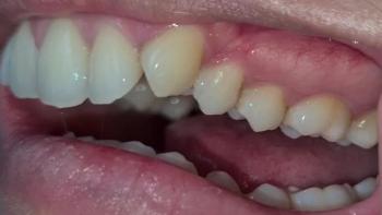
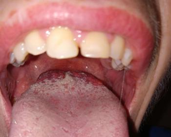
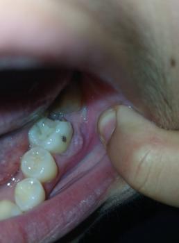
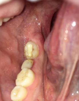
Patients with lower front gum inflammation may also experience tartar buildup, periodontal disease, gum abscesses, or tooth decay. Explore these related clinical case studies.


Other topics:
Other Dental Clinic
 |
 |
 |
 |