Your Smile, Perfected with Precision.

Expert Dental Guide: What Does Dark Discoloration on a Lower Molar Mean
Image:
Severity:
Teeth Problems:
When a Dark Lower Molar Becomes a Serious Dental Problem: An Expert Case Story
A Short, Dynamic Dental Case Story
“Doctor, my back tooth has turned black. Now part of it is breaking apart, and I have a bad taste in my mouth.”
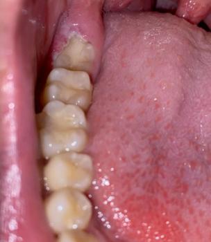
A patient arrived at a dental clinic worried about a dark, damaged lower molar. At first, the tooth appeared to have discoloration. On closer examination, the dentist considered several possible causes, including deep tooth decay, plaque and tartar buildup, and damage to the surrounding gum tissue.
The dentist explained: “A dark tooth does not automatically mean you have a severe infection. However, when discoloration occurs alongside a broken tooth, persistent pain, swelling, or pus, we need to investigate promptly.”
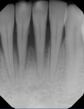
The patient underwent a clinical examination and dental X-rays to determine how much healthy tooth structure remained and whether the pulp, roots, or surrounding bone might be affected.
The treatment plan would depend on the findings. If the tooth remained restorable, treatment might involve removing decay and restoring the tooth. If the pulp was irreversibly damaged or infected, root canal treatment followed by an appropriate restoration might be considered. If the tooth could not be predictably saved, extraction could be necessary.
The dentist emphasized one important lesson: do not wait for a broken or dark tooth to become unbearably painful before seeking dental care. Early assessment can help preserve the tooth when possible and reduce the risk of complications.
Medical Guidance: What Can Cause a Dark Lower Molar?
Dark discoloration around a lower molar can have several explanations:
-
Dental caries (tooth decay): Bacterial activity can progressively destroy tooth structure, leaving brown or black areas and cavities.
-
Plaque and tartar: Accumulated deposits can contribute to discoloration and gum inflammation.
-
Extrinsic staining: Tobacco, coffee, tea, and other staining substances can darken the tooth surface.
-
Pulp damage: Trauma or deep decay may damage the tooth's inner tissue and cause the tooth to darken.
-
Gum or soft-tissue changes: Dark areas on the gums or inner cheek may have causes unrelated to tooth decay and require separate evaluation.
A dentist may use visual examination, dental X-rays, and appropriate pulp and periodontal tests to establish the diagnosis. A photograph alone cannot confirm an abscess, bone infection, or the need for extraction.
Warning Signs of a Possible Dental Infection
Seek urgent dental assessment if a dark or broken molar is accompanied by:
-
Severe or persistent toothache.
-
Swelling of the gum, jaw, or face.
-
Pus, a draining gum bump, or a persistent foul taste.
-
Fever or feeling generally unwell alongside suspected dental infection.
-
Increasing difficulty opening the mouth.
Emergency warning: Rapidly increasing facial or neck swelling, difficulty breathing or swallowing, or swelling that affects the floor of the mouth requires immediate emergency medical care. Do not wait for a routine dental appointment.
Treatment Options for a Broken or Infected Molar
Treatment is determined by the extent of damage, the condition of the tooth's pulp and roots, and the surrounding supporting tissues.
1. Filling or other restoration
Suitable when decay can be removed and enough healthy tooth structure remains to restore function.
2. Root canal treatment and a protective restoration
May be appropriate when the pulp is irreversibly inflamed or infected but the tooth can still be predictably restored.
3. Extraction
May be recommended when the tooth is non-restorable or has a poor prognosis. Replacement options, including a dental implant or bridge, can be discussed after the infection and extraction site have been appropriately managed.
4. Treatment of a dental abscess
The dentist must address the source of infection, which may require drainage, root canal treatment, or extraction. Antibiotics may be indicated in selected cases, especially when there is spreading infection or systemic involvement, but they do not replace definitive dental treatment.
Safe Home Care Before Your Appointment
-
Brush gently with fluoride toothpaste.
-
Rinse gently with warm salt water if comfortable, then spit it out.
-
Choose soft foods and avoid chewing on the damaged tooth.
-
Avoid smoking and limit sugary foods and drinks.
-
Use an over-the-counter pain reliever only if it is safe for you and according to the product label or professional advice.
-
Do not attempt to break, drain, or remove the damaged tooth yourself, and do not place aspirin directly against the gum.
Home care may ease discomfort but cannot repair a severely broken tooth or eliminate an established dental infection.
Frequently Asked Questions
Can a black molar be saved?
Sometimes. The outcome depends on the amount of remaining tooth structure, the condition of the pulp and roots, and the health of the surrounding tissues. An examination and X-rays help determine whether restoration is feasible.
Does a dark tooth always mean infection?
No. Staining, tartar, decay, and changes following trauma can all cause discoloration. Infection must be assessed using the clinical findings rather than color alone.
Can a dental infection spread to the jaw or face?
Yes. An untreated dental infection can spread into surrounding tissues and, in serious cases, cause life-threatening complications. Urgent assessment is important when swelling, fever, or other warning signs develop.
Will antibiotics cure a severely decayed tooth?
Not by themselves. Antibiotics do not repair decay or remove the source of infection. Definitive dental treatment is essential when indicated.
When should I see a dentist?
Arrange an appointment promptly for a dark or broken tooth. Seek same-day urgent dental care if you have significant pain, pus, or swelling, and seek emergency care for breathing or swallowing difficulty or rapidly spreading swelling.
Final Dental Expert Commentary
A dark lower molar deserves attention, particularly when the tooth is crumbling or the surrounding gums are inflamed. The priority is to identify the cause, determine whether the tooth can be saved, and treat any infection before it progresses.
Do not rely on discoloration alone to diagnose a severe infection. A qualified dentist must assess the tooth and surrounding tissues before recommending treatment.
Related case study: Dark Discoloration Near Lower Molar Case Analysis
Medical disclaimer: This educational content is not a substitute for an in-person dental examination, diagnostic imaging, or individualized treatment advice. The case story is illustrative and does not represent a confirmed diagnosis or a documented patient's treatment outcome.
Dark discoloration on a lower molar can range from a harmless surface stain to a sign of tooth decay or internal damage. Because lower molars have deep chewing grooves and multiple surfaces that can be difficult to clean, dark spots may develop in areas where plaque and food debris accumulate. However, color alone cannot determine whether a tooth is healthy, decayed, or infected.
A dental examination helps identify the cause and determine whether the tooth needs professional cleaning, preventive treatment, a filling, root canal treatment, or another procedure.
1. Common Causes of Dark Discoloration on a Lower Molar
Dental caries (tooth decay)
Tooth decay can appear as a brown or black spot in the grooves of a molar or along its sides. As decay progresses, the affected area may become soft, form a cavity, or cause sensitivity and pain. Some cavities cause no noticeable symptoms during their early stages.
Extrinsic stains from food, drinks, and tobacco
Coffee, tea, cola, strongly pigmented foods, and tobacco can contribute to dark deposits on the tooth surface. A professional cleaning may remove external stains, but cleaning cannot repair a cavity or reverse internal tooth discoloration.
Plaque and hardened tartar
Plaque can accumulate around the gumline and between teeth. When plaque hardens into calculus (tartar), it cannot be removed reliably with ordinary brushing and usually requires professional scaling. Tartar may appear yellow, brown, or dark.
An old filling or dental restoration
An existing metal filling or changes around the edges of a restoration may make a molar appear darker. A dentist must assess whether the restoration remains intact and whether decay has developed around it.
Cracked or damaged tooth structure
A crack, fracture, or damaged cusp may allow staining to become more noticeable. Some cracks cause pain when biting or releasing the bite, while others produce few symptoms. Suspected cracks require professional assessment.
Internal discoloration or damage to the dental pulp
A tooth may become gray or dark after an injury or when its pulp—the tissue containing nerves and blood vessels—becomes damaged. In some cases, pulp necrosis (death of the pulp tissue) can occur. A dark tooth alone does not prove that the pulp is dead, so additional examination and testing are necessary.
2. Symptoms That May Accompany a Dark Lower Molar
Pay attention to symptoms in addition to the tooth's color, including:
-
Sensitivity to cold, hot foods, or sweets.
-
Pain when chewing or biting.
-
A visible hole, broken edge, or rough surface.
-
Persistent toothache or pain that occurs spontaneously.
-
Swollen, tender, or bleeding gums near the tooth.
-
A bad taste, pus, or a pimple-like bump on the gum.
-
Swelling of the cheek or jaw.
The absence of pain does not rule out tooth decay or other dental disease.
3. How a Dentist Diagnoses Dark Discoloration
An accurate diagnosis requires more than looking at the color of the tooth. A dentist may:
-
Ask when the discoloration began and whether there has been pain, trauma, sensitivity, or previous dental treatment.
-
Examine the molar, its grooves, the surrounding gum tissue, and any existing fillings or crowns.
-
Clean and dry the tooth when appropriate to assess the affected surface.
-
Take dental X-rays if indicated to evaluate suspected decay between teeth, the extent of a lesion, or changes around the roots.
-
Perform pulp sensibility tests and other clinical tests if internal damage or infection is suspected.
Not every dark spot requires an X-ray, and no single test is sufficient for every case. The dentist combines the findings to establish the most likely diagnosis.
4. Treatment Options for a Dark Lower Molar
Treatment depends on the underlying cause and the extent of any damage.
Professional dental cleaning: Suitable for external stains and tartar when the underlying tooth structure is healthy.
Fluoride treatment and monitoring: Some early, non-cavitated enamel lesions can be managed with fluoride, improved oral hygiene, dietary changes, and follow-up. A formed cavity generally cannot be repaired by brushing or fluoride toothpaste alone.
Dental filling: A dentist may remove decayed tooth structure when indicated and restore the affected area with an appropriate filling material.
Dental crown or other restoration: A molar with extensive structural damage may need a stronger restoration to protect its remaining tooth structure.
Root canal treatment: If the pulp is irreversibly inflamed or infected and the tooth can be saved, root canal treatment may be recommended. The tooth is then restored appropriately.
Extraction: Removal may be considered when a tooth is too extensively damaged to restore predictably or cannot otherwise be retained safely.
Not every dark molar needs a root canal, crown, or extraction. The goal is to preserve healthy tooth structure and choose the least invasive treatment that can predictably restore the tooth's health and function.
5. What You Can Do Before Your Dental Appointment
-
Brush twice daily with fluoride toothpaste and a soft-bristled toothbrush.
-
Clean between your teeth daily without forcing floss into a painful area.
-
Limit frequent sugary snacks and acidic drinks.
-
Chew on the other side if biting on the affected molar causes pain.
-
Avoid scraping the dark area with sharp tools or attempting to drill, file, or bleach it yourself.
-
Arrange a dental examination rather than waiting for pain to develop.
Do not place aspirin directly against the tooth or gum, as it can injure oral tissues. Avoid taking leftover antibiotics without a clinician's advice; antibiotics do not repair tooth decay and are not a substitute for definitive dental treatment.
6. When Is Dark Molar Discoloration an Emergency?
Seek urgent dental care if discoloration occurs with severe or persistent tooth pain, pus, a draining gum lesion, or rapidly worsening symptoms.
Facial or jaw swelling, fever with suspected dental infection, or spreading swelling requires prompt assessment. Difficulty breathing or swallowing, swelling extending toward the eye or neck, or rapidly progressing swelling requires emergency medical care immediately.
A dark spot by itself is not usually a medical emergency, but associated symptoms can change the urgency significantly.
Conclusion
Dark discoloration on a lower molar can result from surface staining, tartar, tooth decay, a previous restoration, trauma, or damage to the dental pulp. Its appearance alone cannot establish the diagnosis. A dentist can examine the tooth, perform appropriate tests, and recommend treatment based on the actual cause. Early assessment can help prevent avoidable damage and preserve the natural tooth whenever possible.
Medical information notice: This article is for general educational purposes and does not replace an individual dental examination or diagnosis.
Dark Discoloration Near Lower Molar and Throat Area Case Analysis
What Is Seen in This Case
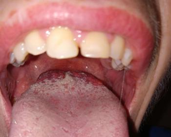
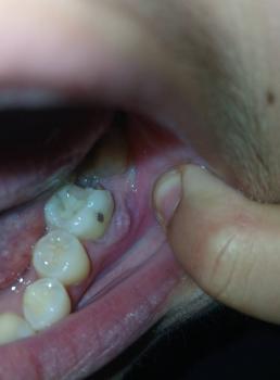
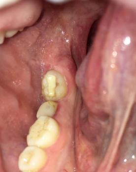
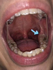
The image shows dark discoloration on the lower back teeth and nearby soft tissue, with the arrow pointing toward the side of the throat and inner cheek area. The lower molars appear heavily stained or decayed, and the surrounding soft tissue looks irritated.
This combination suggests advanced plaque buildup, possible tooth decay, and irritation of nearby oral tissue.
Most Likely Diagnosis
Based on visual examination, the most likely conditions include:
-
Advanced plaque and tartar buildup on lower molars
-
Possible dental caries on back teeth
-
Staining or irritation of soft tissue near the molars
-
Early gum or soft tissue inflammation
-
Risk of bacterial infection spreading to nearby areas
A dental examination and X-ray are required to confirm tooth and bone involvement.
What Causes Dark Discoloration in This Area
Common causes include:
-
Long-term plaque and tartar accumulation
-
Poor oral hygiene around back teeth
-
Smoking or tobacco use
-
Frequent consumption of staining foods or drinks
-
Untreated tooth decay
-
Bacteria irritating nearby soft tissue
Back teeth are harder to clean, making discoloration and decay more likely.
Is This a Serious Problem
This condition can become serious if ignored. If left untreated, it may scale up into:
-
Progressive tooth decay
-
Gum infection or abscess
-
Bad breath that does not improve
-
Pain while chewing
-
Spread of infection to surrounding soft tissue
-
Increased risk of tooth loss
Early treatment helps prevent complications.
Recommended Treatment Process
Initial Assessment (Days 1–3)
-
Dental examination
-
Dental X-ray to check decay depth
-
Evaluation of gum and soft tissue health
Active Treatment Phase (Days 4–7)
Treatment may include:
-
Professional deep cleaning (scaling)
-
Removal of decayed tooth material
-
Dental fillings or further restorative care
-
Treatment of gum or soft tissue inflammation
Healing and Follow-Up Phase (Days 8–14)
-
Reduction in irritation and redness
-
Improvement in oral comfort
-
Planning for long-term oral hygiene maintenance
Expected Healing Time
-
Gum and soft tissue irritation: 5–7 days
-
Recovery after cleaning or minor treatment: up to 14 days
Healing depends on how early treatment begins.
What Happens If Treatment Is Delayed
If delayed beyond 14 days, the condition may worsen and lead to:
-
Severe tooth decay
-
Gum infection or abscess
-
Persistent pain
-
Tooth loss
-
Spread of infection to nearby tissue
-
Higher treatment costs
Home Care While Waiting for Treatment
These steps may help reduce symptoms but do not replace dental care:
-
Brush gently twice daily
-
Floss carefully around back teeth
-
Rinse with warm salt water
-
Avoid smoking and sugary foods
Professional Comment
This case shows significant plaque buildup and possible decay near lower molars, with irritation extending toward nearby soft tissue. Early dental intervention is essential to stop progression and protect both teeth and surrounding oral structures.
Visit a Dental Clinic Near You
For professional evaluation and treatment, visit:
https://cebudentalimplants.com/map-dental-clinic
Related Clinical Cases
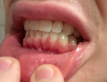
Patients with lower front gum inflammation may also experience tartar buildup, periodontal disease, gum abscesses, or tooth decay. Explore these related clinical case studies.