Healthy Teeth, Happy Life.

Exposed Gum Tissue Around a Lower Molar: Causes, Warning Signs, and Dental Treatment
Image:
Severity:
Teeth Problems:
Exposed Gum Tissue Around a Lower Molar: Causes, Warning Signs, and Dental Treatment
1. Understanding Exposed Gum Tissue Around a Lower Molar
Exposed gum tissue around a lower molar can refer to several different conditions. The gumline may move away from the tooth, exposing part of the root. Alternatively, the gum may appear swollen, irritated, separated from the tooth, or damaged by inflammation or trauma.
A dentist must distinguish between gum recession, periodontal pocket formation, and localized gum swelling because these conditions have different causes and treatments.
Lower molars are important for chewing and maintaining normal bite function. Problems in this region may go unnoticed until a patient develops sensitivity, food trapping, bleeding, discomfort, or persistent bad breath.
A dental professional may examine gum measurements, plaque and tartar deposits, tooth mobility, decay, existing restorations, and the surrounding bone. Dental X-rays may be appropriate when bone loss, root infection, or other underlying problems are suspected.
2. Common Causes of Exposed or Abnormal Gum Tissue
Gum recession: The gum margin moves away from the crown, exposing the root surface. Contributing factors can include periodontal disease, aggressive brushing, thin gum tissue, tooth position, and previous dental treatment.
Gingivitis: Plaque accumulation causes gum inflammation, redness, swelling, and bleeding. Gingivitis generally does not cause permanent attachment or bone loss and may improve with professional cleaning and effective daily oral hygiene.
Periodontitis: This inflammatory condition can damage the supporting tissues and bone around teeth. It may produce periodontal pockets, gum recession, bleeding, persistent bad breath, and eventually loose teeth.
Food impaction or local trauma: Food repeatedly trapped between lower molars, sharp tooth edges, dental restorations, or mechanical irritation can inflame nearby gum tissue.
Tooth decay or infection: Deep decay, a cracked tooth, or an infection near a tooth root can cause localized pain and swelling. A draining gum lesion may indicate an infection and should be examined promptly.
These conditions cannot reliably be distinguished by symptoms alone. Appropriate treatment depends on the clinical findings and the underlying cause.
3. How a Dentist Evaluates and Treats the Condition
An experienced dentist or periodontist evaluates the affected lower molar before recommending treatment. The goals are to control inflammation, preserve the tooth when possible, protect exposed root surfaces, and prevent further damage to supporting tissues.
Step 1: Clinical Gum Examination
The dentist checks for bleeding, recession, tenderness, gum pockets, food impaction, and tooth mobility. This examination helps determine whether the tissue is inflamed, receding, or affected by periodontal damage.
Step 2: Professional Cleaning and Periodontal Treatment
If plaque and tartar contribute to gingivitis or periodontitis, professional cleaning or scaling and root planing may be indicated. Deep cleaning removes deposits from below the gumline and helps control periodontal inflammation.
Step 3: Protecting the Exposed Tooth Root
For sensitivity caused by recession, a dentist may recommend desensitizing toothpaste, fluoride treatment, or another protective treatment. A gum graft may be considered when recession requires root coverage or additional tissue protection.
Gum grafting is not automatically necessary whenever a root is exposed. The appropriate treatment depends on the extent of recession, periodontal health, sensitivity, root-caries risk, and the patient's individual needs.
Step 4: Investigating Deeper Tooth or Bone Problems
If symptoms suggest deep decay, root infection, or bone loss, dental X-rays and additional tests may be needed. Depending on the diagnosis, treatment may involve a filling, root canal treatment, periodontal surgery, or extraction when a tooth cannot be saved.
4. What Patients Can Safely Do at Home
Home care can reduce irritation and help prevent further plaque accumulation, but it cannot replace professional treatment for established periodontitis or a tooth infection.
-
Brush twice daily with a soft-bristled toothbrush and fluoride toothpaste. Use gentle pressure around the affected molar.
-
Clean between the teeth daily using floss or an appropriate interdental cleaner without forcing it into inflamed tissue.
-
Avoid repeatedly poking the gum with toothpicks, fingernails, or sharp objects.
-
Choose comfortable foods if chewing aggravates the area, and avoid foods that repeatedly become trapped around the affected tooth.
-
Arrange a dental appointment if the gumline appears to be changing, sensitivity persists, or bleeding and swelling recur.
Do not attempt to cut away, pull back, or reposition gum tissue yourself. Do not take leftover antibiotics or use antibiotics as a substitute for treating the underlying dental problem.
5. When Exposed Lower-Molar Gum Tissue Needs Urgent Attention
Arrange a dental examination promptly for persistent gum bleeding, worsening sensitivity, recurrent food trapping, a new gum defect, or a tooth that feels loose.
Seek urgent dental care for severe or persistent tooth pain, pus, a draining gum lesion, or localized gum or facial swelling. These symptoms may indicate infection and require prompt treatment.
Seek emergency medical care immediately if you experience difficulty breathing or swallowing, rapidly spreading facial or neck swelling, or swelling that threatens the airway. Fever accompanied by suspected dental infection also warrants urgent assessment, particularly when swelling or worsening pain is present.
A dental infection can spread beyond the original tooth or gum area. Do not wait for a routine appointment if severe symptoms develop.
Common Causes
Gum recession
The gum margin moves away from the tooth, exposing the root. The affected area may become sensitive to cold drinks, sweets, brushing, or touch.
Gingivitis or periodontitis
Plaque accumulation can cause inflammation and bleeding. Advanced periodontitis may destroy the supporting tissues and bone, contributing to gum recession and tooth mobility.
Partially erupted wisdom tooth
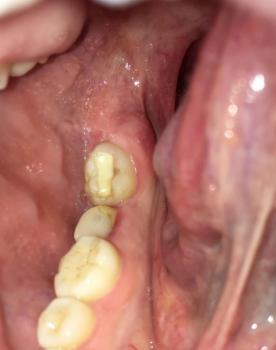
If the affected area is behind the last lower molar, a partially erupted wisdom tooth may trap food and bacteria beneath a gum flap. This can cause a condition called pericoronitis.
Mechanical irritation
Brushing too aggressively, repeated trauma, and certain tooth-position or bite-related factors may contribute to gum damage or recession in susceptible areas.
6. Short Patient Story: A Web Developer With an Exposed Lower Molar
Fictional educational case study — not a real patient record.
Mark, a 38-year-old web developer, spent long hours maintaining websites and fixing Drupal modules. During a busy week, he noticed that the gum around his lower-right molar looked different. Cold water caused mild sensitivity, and food seemed to get trapped between his back teeth more often than before.
At first, Mark assumed that brushing harder would solve the problem. He began scrubbing the affected area more aggressively, but the gum became irritated and occasionally bled.
Concerned that he might lose his tooth, Mark scheduled a dental examination.
The dentist found an area of gum recession and plaque accumulation around the lower molar. Because the appearance alone could not establish the full cause, the dentist measured the periodontal pockets and assessed the tooth for decay and supporting-bone problems.
The dentist explained that exposed root surfaces can become sensitive and are more vulnerable to root decay. Mark was advised to use a soft-bristled toothbrush with gentle technique, improve daily cleaning between his teeth, and follow the recommended professional treatment plan.
After treatment and follow-up, Mark learned that the important goal was not simply to make the gum look normal again. It was to control the underlying cause, protect the tooth, and prevent further damage.
Lesson from Mark's experience: When gum tissue around a lower molar changes, early dental assessment is more useful than guessing at the cause or trying increasingly aggressive home remedies.
7. Frequently Asked Questions
1. Why is the gum around my lower molar pulling away?
Possible causes include periodontal disease, aggressive brushing, naturally thin gum tissue, tooth positioning, and local trauma. A dentist must examine the area to determine the most likely cause.
2. Can exposed gum tissue grow back naturally?
Gum tissue lost through established recession generally does not grow back on its own. Inflamed gum tissue may improve after appropriate treatment if the condition is gingivitis rather than established tissue loss. Some cases of recession can be treated with gum grafting or other root-coverage procedures.
3. Does exposed gum tissue always mean gum disease?
No. Recession may result from brushing trauma, thin gum tissue, tooth position, or other factors. However, periodontal disease is an important possible cause, particularly when bleeding, deep pockets, bone loss, or tooth mobility is present.
4. Can I keep my lower molar if the root is exposed?
Often, yes. An exposed root does not automatically mean a tooth must be extracted. The outlook depends on the amount of supporting bone and tissue, whether decay or infection is present, and the tooth's overall condition.
5. Is gum graft surgery necessary for every exposed root?
No. A dentist or periodontist may recommend monitoring, improved oral hygiene, treatment for periodontal disease, desensitizing products, or other measures. Surgery may be appropriate when root coverage or additional tissue protection is needed.
6. Why does my lower molar hurt when I drink cold water?
Sensitivity may occur when gum recession exposes the root surface. However, tooth decay, a cracked tooth, and other dental conditions can produce similar symptoms. Persistent or worsening pain should be examined rather than automatically attributed to recession.
7. Can mouthwash repair receding gums?
Mouthwash may help control plaque or improve oral hygiene when appropriately selected, but it cannot regenerate gum tissue already lost through recession. It should not replace professional periodontal treatment when indicated.
8. Should I take antibiotics for swollen gum tissue?
Not automatically. Many gum problems require plaque removal, periodontal treatment, or treatment of the affected tooth rather than antibiotics. A dentist must assess whether an infection is present and whether antibiotics are appropriate.
9. How can I prevent further gum recession around my molars?
Brush gently with a soft-bristled toothbrush, clean between your teeth daily, attend recommended dental checkups, and avoid tobacco. If you notice new recession or persistent bleeding, have the area evaluated.
10. When should I see a dentist?
Schedule an appointment for new or progressive recession, persistent sensitivity, recurrent bleeding, or food trapping. Seek urgent care for severe pain, pus, or swelling, and emergency care for difficulty breathing or swallowing.
8. Suggested SEO Additions
Suggested H2 heading: Exposed Gum Tissue Around a Lower Molar: Causes and Treatment Options
Related search phrases:
-
Exposed gum around lower molar
-
Gum recession around back teeth
-
Exposed molar root sensitivity
-
Swollen gum behind lower molar
-
Periodontal treatment for lower molars
Suggested internal links:
-
Your existing Exposed Gum Tissue Around a Lower Molar article.
-
A relevant article on gum inflammation or gingivitis.
-
A relevant article on periodontal disease and bone loss.
-
A relevant article on dental abscess symptoms and treatment.
Suggested meta description:
Learn about exposed gum tissue around a lower molar, including possible causes, gum recession, warning signs, dental treatments, home care, and FAQs.
9. Medical Review and Website Disclaimer
This content is intended for general dental education. It does not diagnose individual conditions or replace an examination, diagnosis, or treatment plan from a licensed dentist.
For clinical accuracy, have a licensed dentist review the content before presenting it as professionally reviewed or dentist-authored. Do not label it medically reviewed by a dentist unless that review has actually occurred.
Suggested reference sources for editorial review:
-
American Dental Association: https://www.ada.org/resources/ada-library/oral-health-topics/periodontitis
-
American Academy of Periodontology: https://www.perio.org/for-patients/periodontal-treatments-and-procedures/surgical-procedures/
-
Cleveland Clinic: https://my.clevelandclinic.org/health/diseases/22753-gum-recession
Exposed Gum Tissue Near Lower Molar Case Analysis (No Protective Skin)
What Is Seen in This Case
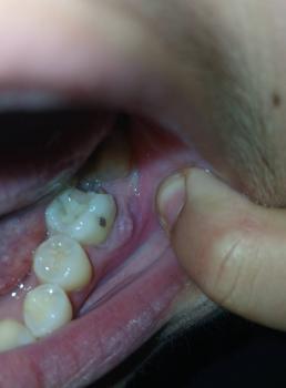
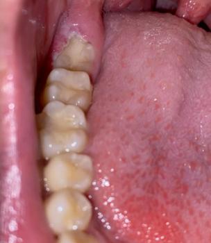
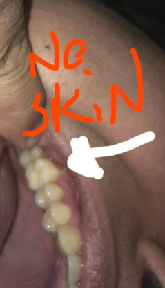
The image shows an area near the lower back teeth where the gum tissue appears thin or missing, exposing the underlying soft tissue. The marked area (“no skin”) suggests loss of normal gum coverage, which can make the area sensitive, sore, and vulnerable to infection.
The nearby teeth appear intact, but the gum condition indicates localized tissue damage or recession.
Most Likely Diagnosis
Based on visual examination, the most likely conditions include:
-
Gum recession near the lower molar
-
Loss of protective gum tissue
-
Localized gum trauma or irritation
-
Early infection risk due to exposed tissue
-
Possible post-extraction or post-inflammation tissue loss
A dental examination is required to determine the cause and depth of tissue involvement.
What Causes Loss of Gum “Skin” in This Area
Common causes include:
-
Aggressive brushing or repeated friction
-
Previous tooth extraction or dental surgery
-
Chronic gum inflammation
-
Food impaction causing repeated irritation
-
Poor oral hygiene leading to tissue breakdown
Once gum tissue is damaged, it does not easily regrow without care.
Is This a Serious Problem
This condition can become serious if ignored. If left untreated, it may scale up into:
-
Persistent pain or sensitivity
-
Infection of the exposed tissue
-
Worsening gum recession
-
Bone exposure in advanced cases
-
Increased risk of tooth loss
-
Difficulty chewing comfortably
Early treatment helps protect the area and prevent progression.
Recommended Treatment Process
Initial Assessment (Days 1–3)
-
Dental examination
-
Evaluation of gum thickness and health
-
Check for infection or trauma source
Active Treatment Phase (Days 4–7)
Treatment may include:
-
Professional cleaning
-
Medication or topical treatment if inflamed
-
Protection of the exposed area
-
Adjustment of brushing technique
Healing and Protection Phase (Days 8–14)
-
Gum tissue irritation should reduce
-
Sensitivity should improve
-
Further treatment planned if tissue loss persists
In some cases, gum grafting or specialist care may be recommended.
Expected Healing Time
-
Mild tissue irritation: 5–7 days
-
Moderate gum damage: up to 14 days
-
Long-term tissue repair depends on severity and treatment
What Happens If Treatment Is Delayed
If delayed beyond 14 days, this condition may worsen and cause:
-
Ongoing pain and sensitivity
-
Gum infection
-
Progressive tissue loss
-
Bone exposure
-
Higher treatment cost
Gum tissue problems should be addressed early.
Home Care While Waiting for Treatment
These steps help reduce irritation but do not replace dental care:
-
Brush gently with a soft toothbrush
-
Avoid touching the area with fingers or tongue
-
Rinse gently with warm salt water
-
Avoid spicy, hot, or hard foods
Professional Comment
This case shows loss of protective gum tissue near a lower molar, making the area vulnerable. Early dental evaluation is important to prevent infection, protect bone, and restore comfort.
Visit a Dental Clinic Near You
For gum evaluation and treatment planning, visit:
https://cebudentalimplants.com/map-dental-clinic
Related Clinical Cases
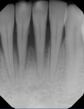
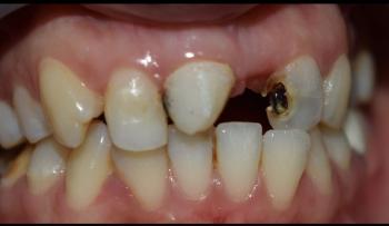
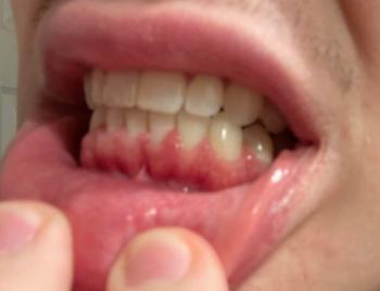
Patients with lower front gum inflammation may also experience tartar buildup, periodontal disease, gum abscesses, or tooth decay. Explore these related clinical case studies.