Your Smile, Our Passion.

Heavy Dental Calculus and Gingivitis: Treatment, Healing Time and FAQs
Image:
Severity:
Teeth Problems:
Clinical Add-On: Heavy Dental Calculus and Gingival Inflammation — Diagnosis, Professional Treatment, and Gum Healing
1. Why Heavy Dental Calculus Requires Professional Treatment
Heavy dental calculus, commonly called tartar, develops when dental plaque accumulates on tooth surfaces and mineralizes over time. It frequently appears along the gumline and behind the lower front teeth, where saliva can contribute to mineral deposition.
Once plaque hardens into calculus, brushing and flossing alone cannot remove it. The rough deposits also make effective plaque control more difficult, allowing bacterial biofilm to persist around the teeth and gingival margins.
From a clinical perspective, the primary concern is not simply the visible amount of tartar. The dentist must determine whether the surrounding tissues show uncomplicated gingivitis or whether periodontal attachment and supporting bone have already been affected.
Gingivitis is inflammation confined to the gingiva and is generally reversible with effective plaque control and professional cleaning. Periodontitis, by contrast, involves loss of the supporting structures of the teeth and requires a more comprehensive periodontal treatment plan.
Clinical principle: The quantity of visible calculus does not independently establish the severity of periodontal disease. Periodontal probing, clinical attachment measurements, and appropriate dental radiographs help determine the extent of damage.
2. Clinical Assessment: What the Dentist Should Examine
In a patient presenting with substantial calculus deposits and inflamed gums, the examination should establish both the distribution of the deposits and the condition of the periodontal tissues.
The assessment should include:
-
Gingival appearance: Evaluate redness, swelling, tenderness, and bleeding.
-
Plaque and calculus distribution: Identify deposits above and below the gumline.
-
Periodontal pocket measurements: Measure the spaces between the teeth and gums at multiple sites around each tooth.
-
Clinical attachment level: Determine whether the periodontal attachment has been lost.
-
Gum recession and tooth mobility: Assess root exposure and the stability of individual teeth.
-
Dental radiographs, when indicated: Evaluate the supporting bone and investigate other suspected dental pathology.
-
Medical and dental history: Review tobacco use, diabetes control, dry mouth, medications, previous periodontal treatment, and oral-hygiene practices.
If a photograph shows redness, swelling, or an elevated area beneath the tongue, that finding must be assessed separately. A photograph cannot reliably determine whether it represents normal anatomy, salivary-duct irritation, infection, or another oral mucosal condition.
A persistent unexplained red or white patch, ulcer, lump, or area of thickening warrants direct dental examination rather than being automatically attributed to calculus.
3. Professional Treatment: Choosing the Correct Approach
A. Routine scaling for calculus and gingivitis
When calculus is present without evidence of periodontitis, professional scaling is generally appropriate. The dentist or dental hygienist removes hardened deposits and plaque from the tooth surfaces, including the gumline.
Polishing may be performed when indicated, but polishing alone does not treat periodontal disease or remove all subgingival calculus.
B. Scaling and root planing for periodontitis
If the examination identifies periodontal pockets, subgingival calculus, and periodontal attachment loss, the dentist may recommend scaling and root planing or other appropriate subgingival instrumentation.
This treatment removes plaque, bacterial biofilm, and calculus from affected tooth and root surfaces. Depending on the extent of disease, treatment may be divided across several appointments.
Not every patient with heavy visible tartar needs deep cleaning. The procedure must be based on the periodontal diagnosis rather than the appearance of the teeth alone.
C. Additional treatment for persistent disease
After initial periodontal treatment, some patients continue to have deep pockets, bleeding, or areas that are difficult to clean. These findings may require further instrumentation, periodontal specialist assessment, or additional treatment.
Periodontal surgery is reserved for selected cases where the clinical findings justify it. It is not an automatic consequence of heavy calculus accumulation.
Antibiotics should not be prescribed routinely for uncomplicated gingivitis or simply because tartar is extensive. They do not replace mechanical removal of calculus and appropriate dental treatment.
4. Expected Healing Timeline After Professional Cleaning
Healing depends on the initial diagnosis, the extent of inflammation, the treatment performed, and the patient's ability to control plaque at home.
First 24–72 hours
Following substantial calculus removal, the gums may feel tender, and temporary tooth sensitivity can occur. Previously inflamed tissue may bleed during cleaning. Mild symptoms should generally improve rather than progressively worsen.
Days 4–7
With effective daily plaque control, gingival redness, swelling, and bleeding may begin to decrease. The patient should continue gentle, thorough brushing rather than avoiding the gumline because it previously bled.
Days 7–14
Patients with plaque-induced gingivitis may notice meaningful improvement during this period. However, the amount of improvement varies, and two weeks should not be treated as a guarantee of complete healing.
Persistent bleeding, swelling, pain, or unexplained oral lesions should be reassessed rather than assumed to be part of normal recovery.
Around 2–6 weeks
A periodontal reassessment may be appropriate after nonsurgical periodontal treatment. The dentist can evaluate residual inflammation, pocket measurements, bleeding on probing, oral-hygiene effectiveness, and the need for further treatment.
Established periodontitis may require ongoing supportive periodontal care even after the initial treatment is completed.
Important distinction: Gum inflammation may resolve, but existing periodontal bone loss does not automatically regenerate after scaling. Long-term treatment aims to control disease, preserve remaining attachment, and reduce the risk of further destruction.
5. Home Care After Calculus Removal
Professional cleaning removes existing calculus, but plaque begins accumulating again soon afterward. Consistent home care is therefore essential.
-
Brush twice daily with fluoride toothpaste and a soft-bristled toothbrush.
-
Clean gently along the gumline without aggressive scrubbing.
-
Clean between the teeth daily using dental floss or appropriately sized interdental brushes.
-
Follow individualized cleaning instructions if periodontal pockets, gum recession, bridges, braces, or implants are present.
-
Avoid tobacco products, which can worsen periodontal outcomes and impair healing.
-
Arrange follow-up cleaning and periodontal maintenance at intervals recommended by the dentist.
An antimicrobial mouth rinse may be appropriate in selected cases, but it should not replace mechanical plaque removal. Chlorhexidine, in particular, should be used only as directed because prolonged use can cause staining and other adverse effects.
Patients should not attempt to scrape heavy calculus off their teeth with household tools or sharp metal instruments. This can injure the gums and damage tooth surfaces.
6. Warning Signs That Require Prompt Assessment
Heavy calculus and gingivitis do not always cause severe pain. Patients should seek prompt dental assessment if they experience persistent bleeding, worsening gum swelling, pus, increasing tooth mobility, pain during chewing, or a localized swelling that does not resolve.
Facial swelling accompanied by fever or rapidly worsening symptoms requires urgent evaluation. Difficulty swallowing, difficulty breathing, swelling beneath the tongue, or rapidly spreading swelling of the floor of the mouth or neck requires emergency medical care.
A suspicious or unexplained oral lesion should also receive direct examination, particularly if it persists for approximately two weeks or longer.
7. Clinical Summary
Heavy dental calculus is a removable local factor that can sustain plaque accumulation and gingival inflammation. The appropriate treatment is professional calculus removal combined with effective daily plaque control.
The decisive clinical question is whether the patient has gingivitis alone or established periodontitis. That distinction determines whether routine scaling is sufficient or whether deeper periodontal treatment and long-term maintenance are necessary.
A realistic treatment plan should prioritize accurate diagnosis, safe mechanical cleaning, individualized home-care instruction, and reassessment of the healing response. No photograph or fixed 14-day timeline can independently establish the final diagnosis or guarantee a specific outcome.
Medical information disclaimer: This educational content is not a substitute for an in-person dental examination, periodontal charting, or diagnostic imaging when indicated.
Frequently Asked Questions About Heavy Dental Calculus and Gingival Inflammation
1. Can heavy dental calculus cause swollen and bleeding gums?
Yes. Plaque accumulation around calculus can sustain gingival inflammation, resulting in redness, swelling, tenderness, and bleeding during brushing or flossing. A dental examination is necessary to determine whether the condition is gingivitis or periodontitis.
2. Can I remove hard tartar at home by brushing harder?
No. Brushing and flossing help control soft plaque, but they cannot reliably remove hardened calculus. Brushing harder may injure the gums and contribute to abrasion. Professional scaling is the appropriate method for removing established deposits.
3. Will my gums heal after scaling?
Gingivitis often improves after professional cleaning and consistent home plaque control. The degree of improvement depends on the original inflammation, the patient's oral hygiene, and other risk factors. Periodontitis may require additional treatment and ongoing maintenance.
4. How long does it take for bleeding gums to stop after professional cleaning?
Bleeding often decreases over several days and may improve substantially within one to two weeks when the underlying cause is controlled. The timeline varies, and persistent or worsening bleeding warrants reassessment.
5. Does heavy tartar automatically mean I have periodontitis?
No. The amount of visible calculus alone cannot confirm periodontitis. A dentist must evaluate periodontal pocket depths, attachment levels, bleeding, recession, tooth mobility, and bone levels when indicated.
6. Is routine scaling enough, or do I need deep cleaning?
Routine scaling may be sufficient when the problem is limited to supragingival calculus and gingivitis. Scaling and root planing or other subgingival instrumentation may be indicated when periodontitis is diagnosed. The decision should follow a periodontal examination.
7. Do antibiotics cure gingivitis caused by tartar?
Antibiotics do not remove hardened calculus and are not routinely needed for uncomplicated plaque-induced gingivitis. Treatment generally focuses on professional mechanical cleaning and daily plaque control. Antibiotics are reserved for selected clinical situations when indicated by a qualified clinician.
8. Why do my teeth feel sensitive after scaling?
Temporary sensitivity may occur when substantial deposits are removed, previously covered root surfaces become exposed, or inflamed tissues begin healing. Sensitivity should be assessed if it is severe, persistent, or associated with other symptoms.
9. Can gum disease cause bad breath?
Yes. Persistent plaque, calculus, gingival inflammation, and periodontal pockets can contribute to bad breath. Other causes include tongue coating, dry mouth, dental decay, and certain medical conditions. Persistent halitosis should be evaluated rather than treated solely with mouthwash.
10. Can untreated gingivitis lead to loose teeth?
Gingivitis alone does not cause the attachment and bone loss characteristic of periodontitis. However, periodontitis can progressively damage the structures supporting the teeth, potentially resulting in mobility and tooth loss. Not everyone with gingivitis develops periodontitis, but persistent inflammation should be addressed.
11. Should I wait 14 days before seeing a dentist?
No. The two-week period is a possible window for monitoring improvement after appropriate treatment, not a reason to delay initial assessment. Arrange a dental appointment when heavy calculus or persistent gum inflammation is present. Seek urgent care for spreading swelling, fever with dental swelling, or difficulty swallowing or breathing.
12. When should I return for follow-up?
The timing depends on the diagnosis and treatment. A patient with uncomplicated gingivitis may need routine follow-up and preventive cleaning, while a patient treated for periodontitis may need a periodontal reassessment and a personalized maintenance schedule. The dentist should determine the interval from the clinical findings.
A dentist would need periodontal probing, bite/occlusal examination, and usually appropriate radiographs to determine bone loss and whether there is an underlying dental infection.
What I can see at 100% view
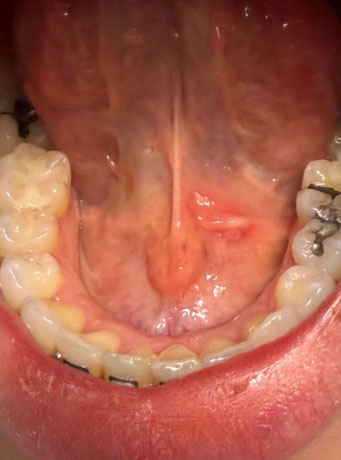
The most obvious finding is substantial hardened calculus/tartar on the tongue-side (lingual) surfaces of the lower front teeth, particularly around the lower incisors. There also appears to be localized redness/inflammation of the soft tissue under the tongue, near the floor-of-mouth/frenulum region.
The image does not by itself establish the severity of periodontal bone loss.
1. Heavy calculus — HIGH confidence
The yellow/brown deposits around the lower anterior teeth look consistent with mature dental calculus rather than simply soft plaque.
This is important because calculus provides a rough surface where plaque can accumulate and can contribute to gingival inflammation.
2. Gingival inflammation — likely
The surrounding gingival tissues appear somewhat erythematous. This could be compatible with gingivitis, but if periodontal pockets and bone loss are present, it could represent periodontitis.
A photograph cannot distinguish those reliably.
3. Floor-of-mouth lesion — needs attention
There is a localized reddish/elevated area beneath the tongue, close to the lingual frenulum.
This could have relatively benign explanations, including irritation or inflammation around normal salivary-duct structures, but the photograph alone cannot determine what it is.
This is the part I would specifically have a dentist/oral-medicine clinician examine.
What I would do clinically
Step 1 — Comprehensive examination
A dentist should document:
- Full periodontal charting
- Pocket depths around every tooth
- Bleeding on probing
- Tooth mobility
- Recession
- Furcation involvement where applicable
- Plaque/calculus levels
- Condition of existing restorations
- Examination of the entire floor of mouth and tongue
- Palpation of the floor of mouth and neck lymph nodes
The ADA recommends a conventional visual and tactile examination for oral mucosal abnormalities rather than relying on photographs alone.
Step 2 — Radiographs if clinically indicated
Depending on the periodontal findings, the dentist may obtain appropriate intraoral radiographs to determine whether there is:
- Horizontal bone loss
- Vertical bone defects
- Calculus below the gumline
- Root abnormalities
- Periapical pathology
- Other hidden dental disease
Radiographs can provide information that simply cannot be obtained from this photograph.
Step 3 — Professional scaling
For the visible calculus, professional removal is appropriate.
If there is only supragingival calculus, routine scaling may be sufficient.
If there is substantial subgingival calculus and periodontal pocketing, the treatment may instead require scaling and root planing/deep periodontal instrumentation, often divided into sections depending on the patient's findings.
If you want everything scaled
I would not recommend trying to remove this amount of calculus yourself with a metal scaler.
A reasonable professional sequence is:
Visit 1
→ Examination + periodontal charting
→ Determine whether radiographs are needed
→ Remove obvious supragingival calculus where appropriate
→ Establish periodontal diagnosis
Visit 2 or subsequent visits
→ Complete periodontal instrumentation/scaling
→ Root-surface debridement where indicated
→ Oral-hygiene instruction
Re-evaluation
→ Check bleeding, inflammation, pocket measurements and tissue response
→ Determine whether additional periodontal treatment is necessary.
What could improve within 14 days?
If the main problem is plaque/calculus-associated gingival inflammation and the irritant is professionally removed, the soft-tissue inflammation can begin improving within days.
However, 14 days does not guarantee complete healing.
A useful way to think about it:
| Finding | Possible response |
|---|---|
| Soft plaque | Improves quickly with effective cleaning |
| Gingival inflammation | Often noticeably improves over 1–2 weeks |
| Heavy calculus | Requires professional removal |
| Deep periodontal pockets | May require additional periodontal treatment |
| Bone loss | Does not simply regenerate in 14 days |
| Local traumatic irritation | May improve within days after the irritant is removed |
| Persistent unexplained mucosal lesion | Requires professional reassessment |
The important 14-day issue
The floor-of-mouth/red area is the finding I would not simply ignore for 14 days.
If that area is new and clearly attributable to irritation, the dentist may choose to remove the suspected irritant and observe the healing response.
But if a red/white lesion, ulcer, lump, thickening, or other unexplained abnormality persists for approximately two weeks, it should be reevaluated. The ADA states that persistent oral abnormalities warrant further investigation and, when appropriate, biopsy/referral.
That does NOT mean this photograph shows cancer. There are many benign causes of oral lesions. It means persistence beyond the expected healing period changes the clinical approach.
What could "scale up" if treatment is delayed?
If the calculus and inflammation are left untreated, the concern is progression from superficial inflammation to periodontal disease in susceptible patients:
Plaque → calculus → gingival inflammation → periodontal pocket formation → attachment loss → bone loss → tooth mobility/tooth loss
But that progression is not inevitable and cannot be predicted from this photograph alone.
Also, antibiotics are not a substitute for removing the dental cause when the problem is localized periodontal/dental disease. ADA guidance emphasizes definitive dental treatment rather than routine antibiotics for most localized dental conditions.
Seek prompt examination sooner than 14 days if
The floor-of-mouth area develops:
- Rapidly increasing swelling
- Significant pain
- Pus
- Fever
- Difficulty swallowing
- Difficulty breathing
- Difficulty moving the tongue
- Neck swelling
- Rapidly worsening symptoms
Those symptoms require urgent clinical assessment.
My overall impression from this image: the heavy lower-anterior calculus is the clearest dental problem, with probable localized gingival inflammation. The under-tongue red/elevated area deserves a separate examination, because its nature cannot be reliably determined from this photograph.
If this is being prepared as a CebuDentalImplants.com dental case, I would label it “Preliminary photographic assessment — not a definitive diagnosis” and recommend periodontal charting + appropriate radiographs + examination of the floor of mouth before assigning the final diagnosis.
Gingival Inflammation: Expert Clinical Guidance
Gingival inflammation, commonly called gingivitis, is an inflammatory response of the gums that is frequently associated with bacterial plaque buildup along and beneath the gumline. When significant dental calculus (tartar) is present, ordinary brushing cannot remove the hardened deposits. Professional dental cleaning is therefore often an important part of controlling the inflammation.
What Dental Experts Look For
A dental professional assessing inflamed gums will typically evaluate several findings rather than relying on appearance alone:
-
Gum redness, swelling, or tenderness
-
Bleeding during gentle periodontal probing
-
Plaque and calculus accumulation
-
Gum pocket depths
-
Gum recession
-
Tooth mobility
-
Bad breath or an unpleasant taste
-
Signs of localized infection
-
Evidence of bone loss on dental X-rays when periodontal disease is suspected
Importantly, gingivitis does not automatically mean that bone has been lost. Bone loss is more characteristic of periodontitis and requires a proper periodontal examination and, when indicated, radiographic assessment.
Why Heavy Calculus Can Keep Gums Inflamed
Dental calculus is hardened mineralized plaque. Once calculus has formed, brushing and flossing cannot reliably remove it.
The rough calculus surface can make plaque retention easier and can contribute to persistent irritation of the gingival tissues. Removing the calculus professionally allows the patient to maintain a cleaner tooth surface at home.
However, calculus is not the only possible cause of gingival inflammation. Other factors may include inadequate plaque control, smoking or tobacco exposure, certain medications, hormonal changes, systemic conditions, poorly fitting dental restorations, and other oral conditions.
What Happens After Professional Cleaning?
Gums do not necessarily return to normal immediately after calculus removal.
A typical improvement pattern may include:
First several days:
Bleeding or tenderness may temporarily remain, particularly when the gums were significantly inflamed before treatment.
Following 1–2 weeks:
With effective plaque control and appropriate professional treatment, redness, swelling, and bleeding may begin to decrease.
Several weeks:
The dental professional can reassess the gums and determine whether inflammation is resolving or whether additional periodontal treatment is necessary.
The exact timeline varies considerably. Severe inflammation, periodontal pockets, smoking, diabetes, poor oral hygiene, and other factors can affect healing.
When Gingivitis May Be More Than Gingivitis
Persistent bleeding or swelling should not simply be assumed to be uncomplicated gingivitis.
If periodontal pockets, gum recession, tooth mobility, or radiographic bone loss are present, the condition may have progressed to periodontitis. Periodontitis can damage the tissues and bone supporting the teeth and may require treatment beyond routine cleaning.
A dentist or periodontist may recommend periodontal probing, dental X-rays, scaling and root planing, improved home care, and periodic periodontal maintenance depending on the findings.
Expert Clinical Perspective
A useful principle in periodontal care is:
Treat the cause, measure the response, and reassess the tissues.
The goal is not simply to make the gums look better after cleaning. The dental team should determine whether inflammation is resolving, whether periodontal pockets remain, and whether there is evidence of attachment or bone loss.
Patients should also understand that professional treatment and home care work together. Even after calculus has been removed, plaque can begin accumulating again. Daily brushing with fluoride toothpaste, cleaning between the teeth, and regular professional dental care are important for maintaining periodontal health.
When to Seek Prompt Dental Care
Arrange a dental evaluation if you have persistent:
-
Gum bleeding
-
Gum swelling
-
Bad breath
-
Pus or drainage around a tooth
-
Receding gums
-
Loose teeth
-
Pain when biting
-
Increasing tooth sensitivity
-
Changes in the position of your teeth
Seek urgent dental or medical care if facial swelling is rapidly increasing, swelling is affecting the eye or neck, you have difficulty breathing or swallowing, or you develop systemic illness such as significant fever.
Frequently Asked Questions
1. Can heavy tartar cause gingival inflammation?
Heavy calculus can contribute to plaque retention and persistent irritation of the gums. However, inflammation has multiple possible causes, so a dental examination is needed to determine the underlying problem.
2. Can brushing remove heavy dental calculus?
No. Brushing can help control plaque and slow additional buildup, but hardened calculus generally requires professional removal.
3. Will my gums stop bleeding immediately after cleaning?
Not necessarily. Inflamed gum tissue may continue bleeding for a period after treatment. Improvement should occur as inflammation is controlled, but persistent or worsening bleeding should be reassessed by a dental professional.
4. Does bleeding gums always mean periodontal disease?
No. Bleeding can occur with gingivitis and other causes of gum inflammation. However, persistent bleeding deserves evaluation because it can also occur with periodontitis.
5. Does gingivitis cause bone loss?
Gingivitis itself is characterized by inflammation without the loss of periodontal attachment that defines periodontitis. If bone loss is suspected, a dentist may perform periodontal measurements and dental X-rays.
6. Can gum inflammation be reversed?
Early gingivitis is generally considered reversible when the causes of inflammation are effectively controlled. Once periodontitis has caused destruction of supporting tissues and bone, however, the damage is not simply reversed by routine brushing and requires professional periodontal management.
7. How long does it take for inflamed gums to improve?
There is no single healing timeline for every patient. Mild inflammation may improve relatively quickly after effective plaque control, while severe inflammation or periodontal disease may require professional treatment and several follow-up visits.
8. Is scaling painful?
Some patients experience sensitivity or discomfort during or after scaling, especially when there is substantial calculus, gum inflammation, exposed root surfaces, or periodontal pockets. Dental professionals can use appropriate techniques and local anesthesia when indicated.
9. Can mouthwash replace professional dental cleaning?
No. Mouthwash may provide an additional oral-hygiene benefit in selected situations, but it does not remove hardened calculus. It should not be considered a substitute for professional assessment and calculus removal.
10. Can gum inflammation return after treatment?
Yes. Plaque can accumulate again after treatment. Consistent daily oral hygiene and recommended periodontal maintenance are important for reducing recurrence.
11. When should I see a periodontist?
A periodontist may be appropriate when there is suspected or confirmed periodontitis, significant periodontal pocketing, gum recession, bone loss, tooth mobility, or inflammation that does not resolve with appropriate initial care.
12. Is severe gum swelling an emergency?
Not always, but severe or rapidly worsening swelling requires prompt evaluation. Facial swelling accompanied by difficulty breathing or swallowing, significant systemic illness, or swelling spreading toward the eye or neck requires urgent medical attention.
Important Medical Disclaimer
This information is for education and does not replace an examination by a dentist or periodontist. The appearance of gums or photographs alone cannot reliably determine the presence or severity of periodontal disease. Diagnosis may require periodontal probing, clinical examination, and dental radiographs when appropriate.
Treatment should be individualized according to the patient's periodontal findings, medical history, medications, oral-hygiene practices, and risk factors.
Related Clinical Cases
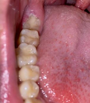
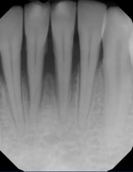
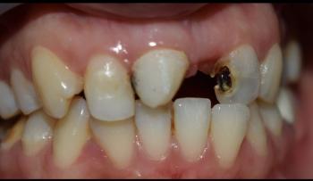
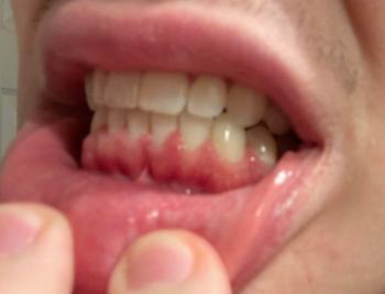
Patients with lower front gum inflammation may also experience tartar buildup, periodontal disease, gum abscesses, or tooth decay. Explore these related clinical case studies.
Other topics:
Other Dental Clinic