From Pain to Perfect — We Care for Your Smile.

Digital Dental X-Rays in the USA: Overview & Advanced Imaging Technology Review
image hero:
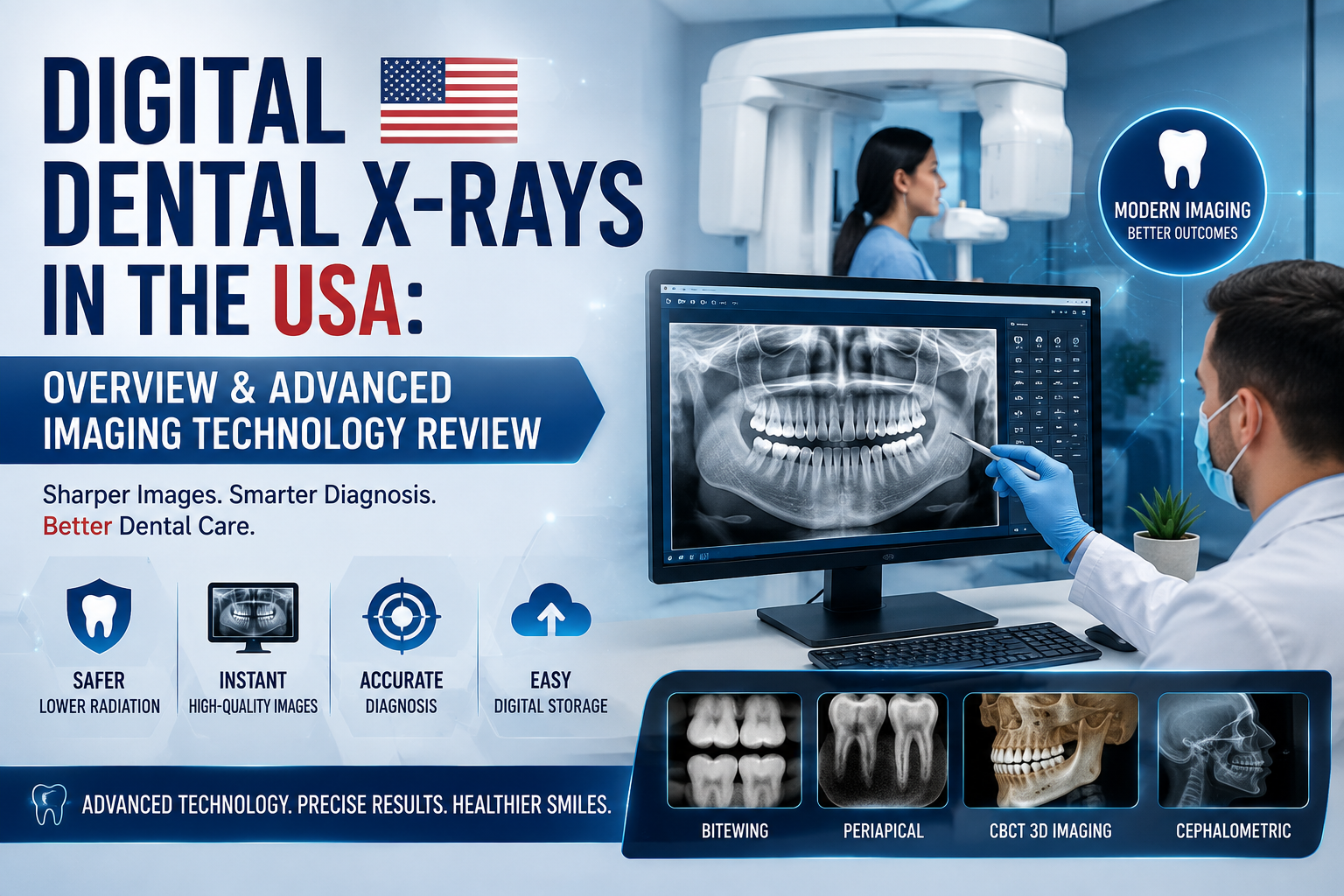
Digital dental radiography is a fundamental component of modern clinical dentistry in the United States. A comprehensive dental examination combines the patient's history, clinical examination, and, when clinically justified, diagnostic imaging.
A dentist cannot see every important structure by looking inside the mouth. Dental radiographs allow clinicians to evaluate structures beneath enamel, between teeth, around tooth roots, and within the supporting jawbone.
The American Dental Association emphasizes that dental X-rays should be prescribed according to the patient's individual clinical needs rather than according to a universal schedule. Factors such as age, oral health status, disease risk, symptoms, previous radiographs, and the purpose of treatment should influence the imaging decision.
1. Why Digital Dental X-Rays Matter
A visual examination can identify many conditions, but some disease processes remain hidden.
Digital radiographs can help the dentist evaluate:
- Interproximal dental caries — decay between teeth that may not be visible during a routine examination.
- Recurrent or secondary caries — possible decay associated with existing restorations.
- Alveolar bone levels — supporting bone changes associated with periodontal disease.
- Periapical pathology — abnormalities around tooth roots.
- Dental abscesses and infection-related changes.
- Impacted or unerupted teeth, including wisdom teeth.
- Root morphology and surrounding anatomy.
- Bone abnormalities and other jaw pathology.
- Treatment-related anatomy for procedures such as endodontics, oral surgery, orthodontics, and implant dentistry.
Radiographs are an adjunct to the clinical examination, not a replacement for it. Diagnosis and treatment planning should not be based on radiographs alone.
2. Major Types of Digital Dental Imaging
Modern American dental offices may use several different imaging technologies depending on the clinical question.
Bitewing Radiographs
Bitewing images primarily display the crowns of the posterior teeth and the interproximal regions.
Common clinical applications include:
- Interproximal caries detection
- Evaluation of existing restorations
- Assessment of marginal alveolar bone levels
- Monitoring patients at appropriate intervals based on individual caries and periodontal risk
Bitewing radiographs are particularly useful when tooth surfaces between adjacent teeth cannot be adequately evaluated by direct visual inspection.
Periapical Radiographs
A periapical radiograph captures the entire tooth from the crown through the root apex, along with the surrounding supporting bone.
It may be selected when evaluating:
- Tooth-root abnormalities
- Periapical infection
- Suspected abscess
- Endodontic disease
- Dental trauma
- Root morphology
- Localized bone pathology
- Post-treatment findings
For a patient presenting with localized tooth pain or swelling, a periapical image may provide information that cannot be obtained from examination of the visible crown alone.
Panoramic Radiography
A panoramic radiograph provides a broad two-dimensional overview of the jaws and dentofacial structures.
Depending on the system and image quality, it can provide information about:
- Upper and lower jaws
- Developing and impacted teeth
- Wisdom teeth
- General dentition
- Condylar regions
- Maxillary sinus regions
- Gross jaw abnormalities
- Overall dental and skeletal relationships
A panoramic image is not a substitute for every intraoral radiograph or CBCT examination. Its broad field of view has limitations involving magnification, distortion, superimposition, and fine-detail visualization.
The appropriate imaging modality depends on the clinical question.
Cone-Beam Computed Tomography
Cone-beam computed tomography, commonly called CBCT, represents an important advancement over conventional two-dimensional dental radiography.
Instead of producing a conventional two-dimensional projection, CBCT generates three-dimensional volumetric information that can be reconstructed in multiple planes.
Common applications include:
- Dental implant treatment planning
- Complex impacted-tooth evaluation
- Selected oral-surgical procedures
- Complex endodontic cases
- Evaluation of maxillofacial anatomy
- Assessment of bone dimensions
- Evaluation of the relationship of teeth or implants to important anatomical structures
For implant dentistry, three-dimensional imaging can provide information about available bone and the relationship of an implant site to critical anatomy.
3. Two-Dimensional X-Ray Compared With CBCT
| Imaging technology | Dimension | Typical strength |
|---|---|---|
| Bitewing | 2-D | Interproximal caries and bone levels |
| Periapical | 2-D | Individual tooth, root and periapical region |
| Panoramic | 2-D | Broad jaw and dentition overview |
| CBCT | 3-D | Complex anatomy and treatment planning |
Key Clinical Principle
More advanced does not automatically mean better for every patient.
If a conventional two-dimensional radiograph can adequately answer the clinical question, exposing the patient to a higher-dose imaging examination simply because CBCT is available may not be justified.
4. Radiation Safety in U.S. Dentistry
Radiographic imaging uses ionizing radiation, so radiation protection is an important part of modern dental practice.
The fundamental principle is ALARA, meaning As Low As Reasonably Achievable.
In practical terms, this means:
Justify the examination, obtain the information needed, and minimize unnecessary exposure.
Important dose-reduction strategies include:
- Using digital image receptors.
- Proper patient positioning.
- Appropriate exposure settings.
- Restricting the X-ray beam to the area of interest.
- Using rectangular collimation when appropriate.
- Avoiding unnecessary repeat images.
- Selecting CBCT only when its additional diagnostic information is justified.
- Following applicable federal, state, and local radiation-safety requirements.
5. Patient Shielding and Modern U.S. Practice
Modern dental radiation protection has evolved significantly.
Current professional guidance no longer recommends routine lead abdominal aprons or thyroid collars for dental radiographic imaging. Instead, modern radiation protection emphasizes appropriate equipment, positioning, beam restriction, exposure optimization, and avoiding unnecessary images.
State and local requirements should still be followed where applicable.
6. How Often Should You Get Dental X-Rays?
There is no single X-ray schedule that applies to every American patient.
Imaging decisions may consider:
- Age
- Previous dental history
- Current oral health condition
- Caries risk
- Periodontal disease risk
- Existing restorations
- Signs and symptoms
- Clinical examination findings
- Previous radiographs
- Whether imaging will change diagnosis or treatment
For example, a patient with active symptoms or significant disease risk may require imaging for a specific diagnostic question, while a low-risk patient with recent adequate images may not need the same examination.
7. Digital X-Ray Technology
Modern digital radiography provides advantages beyond replacing photographic film.
Digital systems can facilitate:
- Rapid image acquisition
- Electronic storage
- Digital transmission
- Image enlargement
- Contrast and brightness adjustment
- Comparison with previous images
- Electronic integration with dental records
- Easier communication between dental professionals
However, digital enhancement does not create anatomical information that was never captured.
An image that is poorly positioned or technically inadequate may still require retaking.
Proper technique and dose optimization therefore remain important.
8. Dental Imaging in Implant Dentistry
Dental implant treatment is one of the areas where advanced imaging can have particularly important clinical value.
A typical implant diagnostic workflow may include:
Clinical examination
↓
Intraoral radiography when indicated
↓
Evaluation of bone and anatomical structures
↓
CBCT when additional three-dimensional information is clinically justified
↓
Implant treatment planning
CBCT can help clinicians evaluate three-dimensional anatomy, including available bone dimensions and the location of important anatomical structures.
However, CBCT should be ordered for a specific clinical purpose rather than simply because three-dimensional imaging is technologically available.
9. Dental X-Rays and Patient Communication
A U.S. dental professional should be able to explain the purpose of an X-ray in understandable language.
For example:
"I recommend this X-ray because there is an area we cannot adequately evaluate during the visual examination. The image will help us determine whether there is decay, infection, bone loss, or another problem and whether treatment is necessary."
This helps patients understand that radiographs are diagnostic tools used when the expected clinical information justifies the examination.
10. Clinical Examination and Imaging
A modern U.S. dental examination should not be thought of simply as looking at the teeth and taking X-rays.
Diagnostic assessment is an integrated process.
Patient History
Medical history, dental history, symptoms, and risk factors.
Extraoral Examination
Face, neck, lymph nodes, temporomandibular joints, and surrounding structures.
Intraoral Examination
Teeth, gingiva, oral mucosa, tongue, palate, and occlusion.
Diagnostic Imaging When Indicated
Bitewing, periapical, panoramic, or CBCT imaging when clinically justified.
Clinical Interpretation
Integration of examination findings and imaging.
Diagnosis
Identification of the disease or condition.
Treatment Planning
Selection of the appropriate intervention.
U.S. Clinical Takeaway
Digital dental X-rays are diagnostic tools that reveal anatomy and disease that may not be visible during a clinical examination.
The modern American approach focuses on patient-specific imaging, radiation optimization, and selecting the imaging modality that answers the clinical question with the least unnecessary exposure.
The core clinical principle is:
Clinical examination first, justify imaging, choose the appropriate modality, optimize exposure, and interpret the image as part of the complete clinical assessment.
Recommended Dental Hub Sections
- Digital Dental X-Rays
- Bitewing X-Rays
- Periapical X-Rays
- Panoramic X-Rays
- CBCT Three-Dimensional Imaging
- Dental Implant Imaging
- Radiation Safety and ALARA
- 2026 ADA and AAOMR Imaging Recommendations
- Dental X-Rays During Pregnancy
- Pediatric Dental Radiography
- Interpreting Dental X-Rays
- X-Ray Versus Clinical Examination
- When CBCT Is Appropriate
- Dental Imaging for Root Canal Treatment
- Dental Imaging for Wisdom Teeth
Related Clinical Cases
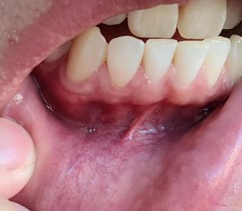
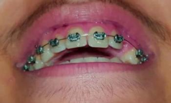
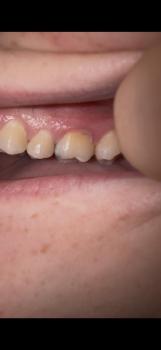
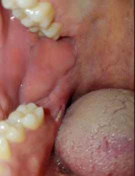
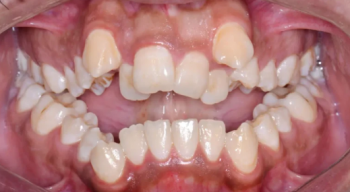
Patients with lower front gum inflammation may also experience tartar buildup, periodontal disease, gum abscesses, or tooth decay. Explore these related clinical case studies.



Other topics:
Other Dental Clinic