We Bring Back the Sparkle in Your Smile.

Clinical Overview & Patient Perspectives: Dental Cysts in the USA
Country:
Dental cysts are abnormal, usually fluid-filled spaces that develop within the jawbone or the tissues surrounding teeth. Common examples include radicular (periapical) cysts, which are associated with teeth that have experienced pulpal inflammation or necrosis, and dentigerous cysts, which develop around the crowns of unerupted teeth.
Many dental cysts grow slowly and may produce few or no symptoms during their early stages. As a result, they are often discovered unexpectedly on routine dental X-rays, panoramic imaging or, when more detailed three-dimensional evaluation is needed, cone-beam computed tomography (CBCT).
What Is a Dental Cyst?
A dental or odontogenic cyst is a pathological cavity involving the jaw or tooth-related tissues. Although many are benign, some can gradually enlarge and affect surrounding structures.
Depending on its type and location, a cyst can potentially cause:
-
Expansion or thinning of the jawbone
-
Displacement of nearby teeth
-
Root resorption
-
Failure of an unerupted tooth to erupt normally
-
Secondary infection
-
In larger lesions, weakening of the jawbone
Some cystic jaw lesions can also resemble other conditions on imaging, which is why a radiographic appearance alone does not always establish the final diagnosis. Histopathological examination may be necessary.
Common Dental Cysts
Radicular Cyst
A radicular cyst, also called a periapical cyst, generally develops around the root tip of a tooth associated with an infected or non-vital pulp.
Treatment depends on the condition of the involved tooth. Root canal treatment may resolve the underlying dental infection, while persistent lesions may require surgical endodontic treatment, extraction or surgical removal of the cystic tissue.
Dentigerous Cyst
A dentigerous cyst develops around the crown of an unerupted tooth. It is particularly associated with impacted third molars, although impacted canines and other teeth can also be involved.
Treatment may include surgical removal of the cyst and the associated tooth. In selected cases, particularly when preservation of an important unerupted tooth is desirable or when the lesion is large, marsupialization or decompression may be considered.
Patient Experiences & Reviews: A USA Perspective
When discussing dental-cyst treatment from a U.S. patient perspective, several recurring themes are important.
1. The “Silent” Discovery
One of the most surprising experiences for patients is learning that they have a jaw cyst despite having little or no pain.
A dentist may discover a suspicious radiolucency during a routine panoramic X-ray or other dental imaging. A cyst can also be discovered while investigating an unerupted or impacted tooth.
This can be unsettling because the patient may feel completely healthy while an abnormality is visible on the X-ray.
2. Anxiety After an Oral-Surgery Referral
Being referred to an oral and maxillofacial surgeon (OMS) can initially create significant anxiety.
Words such as cyst, lesion, jawbone abnormality or tumor can sound frightening to patients. However, the word “lesion” does not automatically mean cancer, and many odontogenic cysts are benign.
A clear explanation of the imaging findings, possible diagnoses and treatment options can help patients understand what is actually being investigated.
3. Imaging Can Be a Major Part of the Diagnosis
A dentist may begin with a periapical or panoramic X-ray. CBCT can provide additional three-dimensional information when the surgeon needs to evaluate the lesion's relationship to teeth, nerves, the maxillary sinus or other anatomical structures.
Imaging helps determine the size and location of a lesion and assists with surgical planning, but the definitive diagnosis may require examination of tissue by a pathologist.
What Happens During Cyst Removal?
Treatment varies according to the cyst type, size, location, associated tooth and potential effect on surrounding structures.
Enucleation
Enucleation involves surgically removing the cystic lining and associated tissue. It is commonly used for appropriate lesions, including many dentigerous cysts.
Depending on the case, the surgeon may also remove an associated tooth or perform additional treatment on the involved tooth.
Marsupialization or Decompression
For selected large cysts, surgeons may choose a more conservative approach.
Marsupialization involves opening the cyst so that it can drain and remain connected to the oral environment. Decompression is another technique used to reduce cyst pressure and gradually decrease lesion size.
These approaches can be particularly useful when immediate complete removal could create substantial surgical risk or when preservation of an important tooth is being considered.
Bone Grafting
A larger cyst can leave a substantial bony cavity after removal. In selected situations, the oral surgeon may recommend a bone graft or another method to help manage the resulting defect.
What Patients Commonly Experience After Surgery
The surgical procedure itself is performed using appropriate anesthesia or sedation based on the patient's needs and the complexity of the case.
After surgery, patients may experience:
-
Facial or intraoral swelling
-
Mild to moderate discomfort
-
Temporary difficulty chewing
-
Dietary restrictions
-
Tenderness around the surgical site
-
Sutures requiring follow-up
-
Gradual bone healing over the following months
Cleveland Clinic notes that many patients can return to normal routines within several days after dentigerous-cyst treatment, although the underlying jawbone may continue healing for months.
Patients should follow the individual postoperative instructions provided by their surgeon because recovery varies according to the size and location of the cyst and the type of procedure performed.
Why Histopathology Matters
One of the most important parts of cyst management is determining exactly what the lesion is.
A lesion that appears to be a particular type of cyst on an X-ray may have a different microscopic diagnosis. For this reason, tissue obtained during surgery may be submitted for histopathological examination.
Research on jaw cystic lesions emphasizes the importance of examining surgical specimens histologically because different lesions can have overlapping clinical and radiographic appearances.
For patients, receiving the pathology report can provide important clarification about what was removed and whether additional treatment or monitoring is appropriate.
Long-Term Follow-Up
Successful removal does not necessarily mean that follow-up is unnecessary.
The oral surgeon or dentist may recommend periodic clinical examinations and radiographic imaging to monitor bone healing and check for recurrence, depending on the type of cyst.
Radiographic follow-up can also demonstrate gradual regeneration of the bone defect. Published cases describe continued radiographic monitoring months after surgery because complete bone remodeling may take substantially longer than the initial soft-tissue recovery.
When Should a Patient Seek Prompt Dental Evaluation?
A person should arrange a dental evaluation if they develop:
-
Persistent jaw swelling
-
A new lump inside the mouth
-
Unexplained tooth displacement
-
A tooth that fails to erupt normally
-
Recurrent swelling around an impacted tooth
-
Persistent pain or pressure
-
Drainage or an unusual taste associated with a tooth
-
Unexplained changes identified on dental X-rays
Urgent medical or dental assessment is particularly important when swelling is accompanied by fever, rapidly increasing facial swelling, difficulty swallowing or breathing, or other signs of a spreading infection.
Key Clinical Takeaways for U.S. Dental Patients
Early detection matters. Because many dental cysts can remain asymptomatic, routine dental examinations and appropriate imaging can identify lesions before they become more extensive.
A cyst does not automatically mean cancer. Many odontogenic cysts are benign, but an accurate diagnosis is important because different jaw lesions can look similar radiographically.
Treatment is individualized. Enucleation, extraction, root canal treatment, marsupialization, decompression, bone grafting or other procedures may be appropriate depending on the specific diagnosis and anatomy.
Histopathology can be essential. Surgical tissue may need microscopic examination to establish the final diagnosis.
Follow-up is part of treatment. Bone healing can continue for months, and some lesions require longer-term clinical and radiographic monitoring.
Patient Perspective: What to Ask Your U.S. Dentist or Oral Surgeon
Before treatment, patients may want to ask:
-
What type of cyst or jaw lesion is suspected?
-
What did my panoramic X-ray or CBCT show?
-
Do I need an oral and maxillofacial surgeon?
-
Is a biopsy or histopathological examination recommended?
-
Does the associated tooth need root canal treatment or extraction?
-
Could the tooth be preserved?
-
Would enucleation, marsupialization or decompression be appropriate?
-
Will the surgical site require bone grafting?
-
How long should I expect swelling and discomfort?
-
How will the area be monitored after treatment?
Bottom Line
Dental cysts can be concerning when first discovered, particularly when the patient has no symptoms and suddenly receives a referral to an oral surgeon. However, many cysts are benign and can be successfully managed when appropriately diagnosed and treated.
For U.S. patients, the most important message is not to ignore an abnormal finding simply because it does not hurt. Proper imaging, specialist evaluation when indicated, appropriate surgical or dental treatment, histopathological confirmation when necessary and long-term follow-up provide the best framework for managing these lesions.
This article is for general educational purposes and does not replace an examination, imaging review or individualized treatment plan from a licensed dentist or oral and maxillofacial surgeon.
Related Clinical Cases
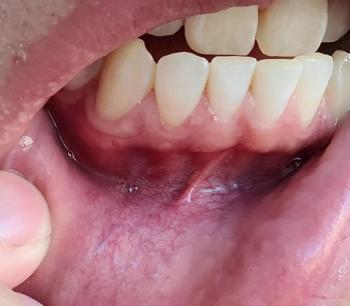
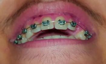
Patients with lower front gum inflammation may also experience tartar buildup, periodontal disease, gum abscesses, or tooth decay. Explore these related clinical case studies.


Other topics:
Other Dental Clinic